Hämostase & Gerinnung
Vollständige Molekularbiologie der antihämostatischen Systeme des medizinischen Blutegels
In einfacher Sprache
Diese Seite erklärt, wie Blutegelspeichel mit dem Gerinnungssystem Ihres Blutes interagiert. Wenn ein Blutegel beißt, liefert sein Speichel den stärksten jemals entdeckten natürlichen Blutverdünner (Hirudin), zusammen mit dutzenden anderer Verbindungen, die das Blut frei fließen lassen. Diese Wissenschaft führte direkt zu Bivalirudin — einem Medikament gegen Herzinfarkt, das jetzt von der American Heart Association als Erstlinientherapie empfohlen wird und jährlich in über einer Million Eingriffen eingesetzt wird.
Where this fits in the bigger picture: Hemostasis mechanism is well-characterised across 440+ catalogued salivary proteins, but the clinical-evidence story is uneven. See the Coverage Map for which conditions actually have RCTs, and the Research Roadmap for what ASH is filling next.
Bildungsinhalt — Mechanismusdiskussion
Einleitung — Hämostase als Grundlage der Blutegeltherapie
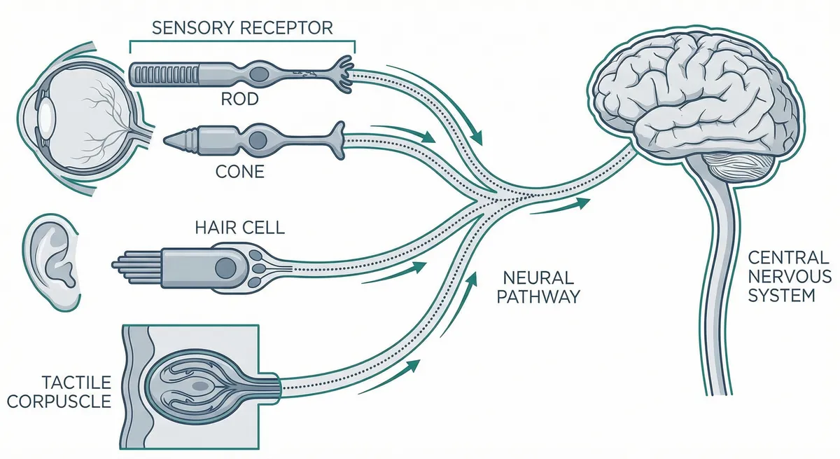
Hemostasis is the body's protective response to disruption of blood vessel integrity — a precisely regulated process that halts hemorrhage while maintaining the fluidity of circulating blood. It is governed by blood flow characteristics, vessel wall structural integrity, endothelial cell functional state, platelets (thrombocytes), and an array of plasma proteins constituting the coagulation and fibrinolytic cascades. Das Verständnis hemostasis at the molecular level is essential to understanding why hirudotherapy is effective: the medicinal leech has evolved the most thorough anti-hemostatic system in nature, with at least 14 distinct compounds that simultaneously target every major step in the hemostatic cascade.
Hemostasis comprises two interdependent arms: the platelet-vascular component (primary hemostasis), which provides rapid initial sealing of the vascular breach, and the plasma coagulation component (secondary hemostasis), which generates fibrin to reinforce and stabilize the platelet plug. Linked to these systems are the kallikrein-kinin system, which initiates the intrinsic pathway, and the fibrinolytic system, which regulates gradual dissolution of the fibrin clot. Jede der these systems represents a molecular target for one or more specific SDS-Komponenten.
Primäre Hämostase
Platelet adhesion and aggregation at the site of vascular injury. Targeted by calin, saratin, decorsin, apyrase, PAF inhibitor, and destabilase.
Sekundäre Hämostase
Coagulation cascade generating thrombin and fibrin. Targeted by hirudin (thrombin), antistasin (factor Xa), kallikrein inhibitor (intrinsic pathway), and ghilanten (factor XIIIa).
Fibrinolyse
Clot dissolution and remodeling. Targeted by destabilase-M (isopeptide bond cleavage) and LCI/TAFI inhibitor (maintains plasminogen binding sites on fibrin surface).
Der mikrovaskuläre Kontext — Wo Blutegeltherapie wirkt
The intradermal vascular bed — where the medicinal leech delivers its SDS — consists almost entirely of microscopic-diameter Blutgefäße. The fundamental building block is the microhemocirculatory unit (Chernukh & Frolov, 1982), comprising the smallest arterial vessels, exchange vessels (capillaries), and the smallest venous vessels. Blood flow velocity in arterioles averages 1.5 mm/s, in capillaries 0.74 mm/s, in venules 0.66 mm/s, and in arteriovenular shunts 1.37 mm/s — compared with 210 mm/s in the aorta. The hematocrit in the microcirculation is typically 2-3 times lower than the systemic hematocrit.
Clinical Significance of Microvascular Delivery
In reconstructive microsurgery, restored arterial inflow may function adequately while venous return remains compromised due to the technical difficulty of venous microanastomosis. The resulting venous congestion causes blood to stagnate in the capillary and postcapillary venular beds, where it rapidly coagulates. By delivering SDS directly into this congested environment — where calin, hirudin, and vasodilatory components achieve high local concentrations — the medicinal leech provides decongestive drainage that no systemic anticoagulant can replicate. The prolonged post-bite bleeding, sustained by calin's blockade of platelet adhesion to collagen, provides continued passive drainage for 8 to 48 hours after der Blutegel detaches.
Vaskuläres Endothel — Das größte endokrine Organ
The endothelium is the largest endocrine organ in the body, weighing approximately 1 kg in a 70-kg adult and covering a surface area of approximately 4,000-7,000 square meters. Endothelial cells synthesize, store, and release a vast array of biologically active substances that regulate vascular tone, cell growth, inflammation, thrombosis, and fibrinolysis. Das Verständnis endothelial function is essential because SDS modulates the hemostatic response at the vascular level.
Anticoagulant Endothelium (Normal)
- • Prostacyclin (PGI2): antiaggregant + vasodilator via cAMP elevation
- • Nitric oxide (NO): inhibits platelet activation/aggregation via cGMP
- • Heparan sulfate proteoglycans: accelerate AT-III 1000-fold
- • Thrombomodulin: converts thrombin to anticoagulant (protein C pathway)
- • TFPI: shuts down initiation phase
- • tPA: constitutively secreted plasminogen activator
- • Ecto-apyrase CD39: degrades ADP to limit platelet activation
Procoagulant Endothelium (Activated)
- • Tissue factor expression: triggers extrinsic pathway
- • P-selectin and E-selectin: leukocyte adhesion
- • PAI-1 upregulation: suppresses fibrinolysis
- • tPA downregulation: reduced plasminogen activation
- • Thrombomodulin downregulation: impaired protein C pathway
- • vWF release: promotes platelet adhesion
- • IL-6, IL-8, chemokines: inflammatory amplification
Prostacyclin-Thromboxane A2 Balance
Both prostacyclin and thromboxane A2 are terminal metabolites of arachidonic acid. Prostacyclin activates adenylate cyclase in the platelet membrane, elevating cyclic AMP, reducing cytoplasmic calcium, and decreasing platelet aggregability. Thromboxane A2, acting through its specific receptors, conversely reduces cAMP and stimulates aggregation. This balance is a principal mechanism by welche die endothelium maintains its athrombogenic surface. SDS contains prostacyclin analogs (6-keto-PGF1-alpha) that supplement endogenous prostacyclin production and may help restore the antithrombotic balance at sites of endothelial damage.
Endothelial Protease Receptors
The endothelium expresses protease receptors that both activate hemostasis and maintain blood fluidity (adapted from Preissner, 2000):
| Protease Ligand | Rezeptor | Cellular Localization | Funktion |
|---|---|---|---|
| Factor VII/VIIa | Tissue factor (TF) | Monocytes, endothelium (activated), adventitial cells | Initiation of coagulation via factor Xa generation |
| Faktor Xa | EPR-1 / Factor Va-phospholipid complex | Endothelium, platelets, monocytes | Thrombin generation via prothrombinase assembly |
| Thrombin | PAR-1, PAR-3, PAR-4 | Endothelium, platelets, smooth muscle, fibroblasts, neurons | G-protein signaling: vascular permeability, cytokine release, cell proliferation |
| Thrombin | Thrombomodulin | Endothelium | Protein C anticoagulant pathway activation; TAFI activation; switches thrombin to anticoagulant |
| Activated Protein C (APC) | Protein S / phospholipid | Platelets, endothelium | Inactivation of factors Va and VIIIa; limits thrombin generation |
Endothelial Damage and the Procoagulant Shift
Inflammatory cytokines (IL-1, TNF-alpha, IFN-gamma) and bacterial endotoxins transform endothelium from thromboresistant to procoagulant and proinflammatory. This procoagulant state is precisely the pathological condition that SDS-Komponenten are evolved to counteract. SDS delivers anti-inflammatory components (eglins, bdellins) and anticoagulant molecules (hirudin, antistasin) into the microenvironment of activated, procoagulant endothelium. The convergent model of hemostasis (Yong & Toh, 2023) suggests that this endothelial activation is not merely a coagulation event but an integrated defense response simultaneously activating innate immune pathways — and SGS's pharmacological breadth is consistent with the evolutionary pressure to counteract this convergent response in its entirety.
Endothelial Regulation of Fibrinolysis
Endothelial cells synthesize three critical regulators of fibrinolysis: urokinase (a polyfunctional cellular regulator), tissue plasminogen activator (tPA) (the only protease continuously secreted in active form), and plasminogen activator inhibitor-1 (PAI-1). The balance between tPA and PAI-1 determines local fibrinolytic capacity. Under inflammatory conditions, PAI-1 is upregulated while tPA is downregulated, creating a fibrinolysis-resistant state that favors thrombus persistence. SDS addresses this through the LCI/TAFI inhibitor, which maintains plasminogen binding sites on the fibrin surface regardless of the endothelial fibrinolytic balance.
Primäre Hämostase — Thrombozytenadhäsion, -aktivierung & -aggregation
When endothelial integrity is disrupted and the underlying smooth muscle cells and extrazelluläre Matrix are exposed, the platelet-vascular component — the phylogenetically most ancient arm of hemostasis — is activated first. While endothelial cells are athrombogenic, the subendothelial surface is highly adhesive to platelets owing to exposed collagen and von Willebrand factor (vWF). Other matrix proteins — fibronectin and laminin — are similarly adhesive. Platelet functions in hemostasis are determined by their ability to adhere to the subendothelial surface, to form aggregates by adhering to one another, and to secrete biologically active compounds from intracellular granules upon activation.
Platelet Receptor Systems
The majority of platelet receptors belong to the integrin class — heterodimeric glycoproteins composed of alpha (130-200 kDa) and beta (90-130 kDa) subunits. Each receptor is targeted by specific SDS-Komponenten:
| Rezeptor | Copies/Platelet | Ligands | Funktion | SDS Inhibitor | Pharma-Analogon |
|---|---|---|---|---|---|
| GP IIb/IIIa (integrin alphaIIbbeta3) | 50,000-80,000/platelet | Fibrinogen, vWF | Final common pathway of aggregation; bridges adjacent platelets | Decorsin (RGD peptide) | Abciximab, eptifibatide, tirofiban |
| GP Ib-V-IX complex (GP Ib-alpha) | ~25,000/platelet | Immobilized vWF | Initial platelet tethering under high shear; primary adhesion receptor | Saratin (indirect — blocks vWF-collagen) | Keine zugelassen |
| GPVI | ~5,000/platelet | Collagen | Major signaling receptor for collagen; activates PLC-gamma2 | Calin (blocks collagen surface) | Revacept (preclinical) |
| Integrin alpha2beta1 (GP Ia/IIa) | ~2,000/platelet | Collagen | Secondary adhesion to collagen; reinforces GPVI signaling | Calin (blocks collagen surface) | Keine |
| PAR-1 (thrombin receptor) | ~1,000-2,000/platelet | Thrombin | Primary thrombin receptor on human platelets; G-protein coupled | Hirudin (blocks thrombin) | Vorapaxar (Zontivity) |
| PAR-4 (thrombin receptor) | Variabel | Thrombin | Secondary thrombin receptor; lower affinity than PAR-1; sustained signaling | Hirudin (blocks thrombin) | Keine zugelassen |
| P2Y1 (ADP receptor) | Variabel | ADP | Initiates shape change and transient aggregation | Apyrase (degrades ADP) | Keine selektiv |
| P2Y12 (ADP receptor) | Variabel | ADP | Amplifies and sustains aggregation; couples to Gi | Apyrase (degrades ADP) | Clopidogrel, prasugrel, ticagrelor |
| Alpha2-adrenergic | Variabel | Epinephrine | Potentiates aggregation response to other agonists | Keine identifiziert | Keine antithrombozytär |
GP IIb/IIIa — The Final Common Pathway
Among platelet receptors, integrin alphaIIbbeta3 (GP IIb/IIIa) holds the leading role, present in 50,000-80,000 copies per cell. On resting platelet membranes, GP IIb/IIIa is weakly activated and does not interact with its ligands — fibrinogen and vWF — which mediate platelet bridging during aggregation. Each agonist (thrombin, ADP, epinephrine, thromboxane A2) engages its specific receptor, and the signal transduction cascade culminates in conformational "opening" of GP IIb/IIIa. Aggregation is completed by bridging bonds between adhesive proteins (fibrinogen, vWF) and activated GP IIb/IIIa on adjacent platelets.
von Willebrand Factor — Shear-Dependent Adhesion
Under static conditions or at low shear rates, direct platelet-collagen interaction (via GPVI and alpha2beta1) suffices for subendothelial adhesion. Under conditions of high shear stress — as in stenotic arteries or arterioles — direct interaction alone is insufficient, and platelet binding to collagen requires additional mediation through vWF (Saelman et al., 1994). The GP Ib-alpha receptor initiates primary contact between platelets and vWF at the vascular wall, after which secondary adhesion and aggregation proceed via GP IIb/IIIa. This shear-rate dependence has direct implications for leech therapy: in the microcirculatory bed where leeches feed (low shear), direct platelet-collagen adhesion predominates — making calin's collagen-binding activity the primary anti-adhesive mechanism.
SDS Anti-Adhesive Components
Calin — Principal Anti-Adhesive
MW ~65 kDa. First evidence for platelet adhesion inhibition obtained by Baskova et al. (1984, 1987): SDS inhibited total platelet adhesion to collagen types I, II, III by 85-87%; initial attachment by 70-80%; spreading by 100%. Type IV collagen pretreated with SDS and thoroughly washed continued to block adhesion (85% attachment inhibition, 100% spreading inhibition), demonstrating that SDS binds to collagen rather than to platelets. Isolated and named by Munro, Jones & Sawyer (1991). Blocks vWF binding to collagen under high shear with IC50 ~0.3 nM (Harsfalvi et al., 1995). Calin is the molecular basis for prolonged post-bite bleeding (4 to 24 hours) that provides therapeutic decongestive drainage in microsurgery.
Saratin — vWF-Collagen Inhibitor
MW ~12 kDa. Isolated by Barnes et al. (2001). Specifically inhibits the vWF-collagen interaction. At low concentrations (high-affinity binding site saturation), blocks platelet adhesion at high shear without affecting collagen-induced aggregation. At high concentrations (low-affinity site saturation), also inhibits collagen-stimulated aggregation. Shear-rate dependence consistent with targeting arterial thrombosis. Rekombinant saratin has shown efficacy in animal models of carotid artery injury — potential antithrombotic for settings where conventional antiplatelet drugs are insufficient.
Decorsin — GP IIb/IIIa Antagonist
MW ~4.4 kDa (39 aa). Isolated from Macrobdella decora (Seymour et al., 1990). Contains Arg-Gly-Asp (RGD) motif enabling direct binding to activated GP IIb/IIIa. IC50 ~100 nM for ADP-induced aggregation. Competes with fibrinogen and vWF for integrin binding, making it a potent agonist-independent aggregation inhibitor. Ornatin (41 aa, from Placobdella ornata) shares the RGD motif — convergent molecular evolution in two distantly related Blutegelart driven by obligate hematophagy. Three FDA-approved GP IIb/IIIa antagonists (abciximab, eptifibatide, tirofiban) target the same receptor.
Apyrase — ADP Degradation
MW 45 kDa (low-MW form) / 400 kDa (high-MW form). Identified by Rigbi, Levy, Eldor et al. (1987). Hydrolyzes ATP to ADP and inorganic phosphate, analogous to endothelial ecto-apyrase CD39. ADP from damaged red blood cells and activated platelet dense granules is a key amplification signal — apyrase interrupts this amplification loop. Unlike P2Y12 antagonists (clopidogrel, prasugrel, ticagrelor) which block one ADP receptor subtype, apyrase eliminates the ADP signal for all receptor subtypes (P2Y1 and P2Y12) simultaneously. P2Y12 antagonists generate peak annual sales exceeding $9 billion.
Evidence: SDS Antiplatelet Studies
| Studie | Design | Population (n=) | Intervention | Primäres Outcome | Ergebnis |
|---|---|---|---|---|---|
| Baskova et al. 1984 | In-vitro-Adhäsionsassay | Human platelets on collagen types I, II, III (n=n.v.) | SDS treatment of collagen-coated surfaces | Platelet adhesion and spreading inhibition | 85-87% inhibition of total platelet adhesion; 70-80% inhibition of initial attachment; 100% inhibition of spreading First evidence that SDS inhibits platelet adhesion; effect independent of collagen type |
| Baskova et al. 1987 | In-vitro-Bindungsstudie | Type IV collagen pretreated with SGS (n=n.v.) | SDS pretreatment of collagen followed by thorough washing | Residual platelet adhesion inhibition | 85% inhibition of initial attachment; 100% inhibition of spreading persisted after washing Demonstrated SDS binds to collagen rather than platelets — key mechanistic insight |
| Munro, Jones & Sawyer 1991 | Proteinisolierung | H. medicinalis SGS (n=n.v.) | Isolation and partial purification of platelet adhesion inhibitor | Identification of calin | Isolated ~65 kDa protein (calin) that blocks platelet adhesion to collagen and vWF binding to collagen Named calin; blocks prolonged post-bite bleeding mechanism |
| Deckmyn et al. 1995 | In-vivo-Tiermodell | Hamster thrombosis model (n=n.v.) | Calin administration | Prevention of platelet-rich thrombi formation | Calin prevented formation of platelet-rich thrombi in hamsters First in vivo demonstration of calin antithrombotic activity |
| Barnes et al. 2001 | Proteincharakterisierung | Saratin from H. medicinalis SGS (n=n.v.) | Characterization of vWF-collagen interaction inhibition at varying shear rates | Shear-dependent antiplatelet activity | 12 kDa protein; blocks platelet adhesion at high shear (low concentration) and collagen-induced aggregation (high concentration) Shear-rate dependence consistent with targeting arterial thrombosis where vWF-mediated tethering is essential |
| Seymour et al. 1990 | Proteinisolierung und -charakterisierung | Macrobdella decora salivary extract (n=n.v.) | Isolation and functional characterization of decorsin | GP IIb/IIIa antagonism via RGD motif | 39-amino-acid RGD peptide; IC50 ~100 nM for ADP-induced platelet aggregation; MW ~4.4 kDa Leech-derived GP IIb/IIIa antagonist; convergent evolution with snake venom disintegrins |
| Baskova et al. 2000 | In-vitro-Aggregationsassay | Human blood platelets with purified destabilase (n=n.v.) | Destabilase incubation with platelets stimulated by various agonists | Inhibition of platelet aggregation | 100% inhibition of spontaneous aggregation; 63% inhibition of ADP-induced (5 uM); 50% inhibition of PAF-induced; 65% inhibition of collagen-induced Destabilase inhibits platelet aggregation via membrane surface interaction, not adenylate cyclase activation |
Evolutionary Multi-Target Strategy
Die Gerinnungskaskade — Von klassischen zu konvergenten Modellen
Das Verständnis hämostatischer Mechanismen hat eine grundlegende Transformation erfahren, seit die klassische Kaskade von Davie & Ratnoff (1964) und Macfarlane (1964) beschrieben wurde. Drei zunehmend vollständigere Modelle — Kaskaden-, Zell-basiertes und konvergentes Modell — beschreiben denselben biologischen Prozess. Alle drei sind relevant für das Verständnis, wie SDS-Komponenten mit der Gerinnung interagieren.
Vollständige Gerinnungsfaktor-Referenz
Die meisten Gerinnungsproteine werden mit römischen Zahlen bezeichnet (Reihenfolge der Entdeckung). Der Buchstabe „a" kennzeichnet aktive Formen, die in den meisten Fällen Serinproteinasen sind. Die Faktoren II, VII, IX und X (der Prothrombinkomplex) werden in der Leber unter Vitamin-K-Kontrolle synthetisiert und enthalten Gamma-Carboxyglutaminsäure- (Gla-) Reste, die die kalziumabhängige Bindung an Phospholipidmembranen vermitteln:
| Faktor | Name | MG | Plasmakonzentration | Halbwertszeit | Funktion |
|---|---|---|---|---|---|
| I | Fibrinogen | 340 kDa | 2-4 g/L | 4–5 Tage | Fibrin-Vorläufer; durch Thrombin in Fibrin-Monomer umgewandelt |
| II | Prothrombin | 72 kDa | 100 ug/mL | 60–72 h | Thrombin-Vorläufer; Gla-Domäne bindet Phospholipidoberflächen über Ca²⁺ |
| III | Gewebefaktor (TF) | 47 kDa | n. v. (membrangebunden) | n.v. | Initiator des extrinsischen Signalwegs; Rezeptor für Faktor VII/VIIa |
| IV | Kalziumionen (Ca²⁺) | 40 Da | 2.2-2.6 mmol/L | n.v. | Essenzieller Kofaktor für Gla-Domänen-Phospholipid-Bindung und Protease-Komplexzusammenbau |
| V | Proakzelerin (labiler Faktor) | 330 kDa | 7 ug/mL | 12–36 h | Kofaktor für Faktor Xa im Prothrombinasekomplex; durch Thrombin aktiviert |
| VII | Proconvertin | 50 kDa | 0.5 ug/mL | 4–6 h | Serinprotease; bindet TF zur Bildung des TF/VIIa-Komplexes; initiiert Gerinnung |
| VIII | Antihämophiler Faktor A | 285 kDa | 0.1 ug/mL | 8–12 h | Kofaktor für Faktor IXa im Tenase-Komplex; Mangel verursacht Hämophilie A |
| IX | Christmas-Faktor (antihämophiler Faktor B) | 57 kDa | 5 ug/mL | 18–24 h | Serinprotease im Tenase-Komplex; Mangel verursacht Hämophilie B |
| X | Stuart-Prower-Faktor | 59 kDa | 10 ug/mL | 40–45 h | Serinprotease; Konvergenzpunkt der intrinsischen und extrinsischen Signalwege; Ziel von Antistasin |
| XI | Plasma-Thromboplastin-Vorläufer | 160 kDa | 5 ug/mL | 40–80 h | Serinprotease; durch Thrombin auf Thrombozytenoberfläche aktiviert (Zell-basiertes Modell) |
| XII | Hageman-Faktor | 80 kDa | 30 ug/mL | 50–70 h | Kontaktaktivierung; für physiologische Hämostase nicht erforderlich (Mangel verursacht keine Blutungen) |
| XIII | Fibrin-stabilisierender Faktor (Transglutaminase) | 320 kDa | 10 ug/mL | 9–12 Tage | Transglutaminase; quervernetzt Fibrin über ε-(γ-Glu)-Lys-Isopeptidbindungen; Ziel von Ghilanten |
| PK | Präkallikrein (Fletcher-Faktor) | 85 kDa | 50 ug/mL | n.v. | Kontaktaktivierung; durch Faktor XIIa zu Kallikrein aktiviert; durch SDS gehemmt |
| HMWK | Hochmolekulares Kininogen (Fitzgerald-Faktor) | 120 kDa | 70 ug/mL | 6,5 Tage | Kofaktor für Kontaktaktivierung; zellulärer Rezeptor für Faktor XI und Präkallikrein |
Der extrinsische Signalweg (Initiation)
Der Auslöser ist der Gewebefaktor (TF), ein 47-kDa transmembranes Glykoprotein, das konstitutiv auf adventitialen Fibroblasten, glatten Muskelzellen und Perizyten exprimiert wird — aber normalerweise auf Zellen abwesend ist, die mit fließendem Blut in Kontakt stehen. Bei Gefäßverletzung bindet TF an Faktor VII/VIIa und bildet einen Komplex, der Faktor X zu Xa und Faktor IX zu IXa aktiviert. Alle Reaktionen finden auf Phospholipidoberflächen in Gegenwart von Kalziumionen statt. TF ist auch ein Mitglied der Zytokinrezeptor-Superfamilie, das intrazelluläre Signalwege über PAR-2 aktiviert und Zellüberleben, Angiogenese und Entzündung fördert.
SDS und der extrinsische Signalweg
The Intrinsic Pathway (Contact Activation)
Activation involves contact-phase proteins (factors XI and XII) and kallikrein-kinin system components: prekallikrein (PK) and high-molecular-weight kininogen (HMWK). PK and factor XI circulate as a complex with HMWK. Binding of HMWK to the endothelial cell surface leads to PK activation to kallikrein, which activates factor XII to XIIa, which activates factor XI to XIa. On the activated platelet surface, factor X is activated at a rate 50- to 100-fold greater than by the TF/VIIa complex. Factor Xa assembles with factor Va in the prothrombinase complex, and prothrombin activation on the platelet surface is amplified more than 200,000-fold.
SDS substantially prolongs the recalcification time of plasma and blocks plasma kallikrein activity through irreversible inhibition (Baskova et al., 1988, 1992), measured using chromogenic substrate S-2302 (D-Pro-Phe-Arg-pNA). The kallikrein inhibitory activity amounts to 14 units per milligram of SDS protein. By inhibiting kallikrein, SDS prevents not only intrinsic pathway activation but also bradykinin generation — kinins enhance pain perception, and leech kininases diminish the pain-inducing effect of bradykinin, an adaptive mechanism for protecting der Wirt from pain during feeding.
The Common Pathway
Factor Xa, regardless of its origin (intrinsic or extrinsic), assembles with factor Va on cell surfaces in the presence of calcium to form the prothrombinase complex, which converts prothrombin to thrombin. Thrombin then cleaves fibrinopeptides A and B from fibrinogen (340 kDa; three pairs of non-identical polypeptide chains: 2-alpha, 2-beta, 2-gamma), generating fibrin monomers that spontaneously polymerize through non-covalent interactions (hydrogen bonds, electrostatic forces between E and D domains). This unstabilized fibrin has limited mechanical strength.
The critical transition to stabilized fibrin is catalyzed by factor XIIIa (transglutaminase), which forms covalent epsilon-(gamma-Glu)-Lys isopeptide bonds between gamma chains (gamma-gamma cross-links, forming within minutes) and between alpha chains (alpha-alpha polymer cross-links, accumulating over hours). This two-stage cross-linking is directly relevant to destabilase-M, which targets these isopeptide bonds. The progressive increase in cross-linking density explains why destabilase's thrombolytic effect increases with stabilization degree — a counterintuitive relationship that distinguishes it from all conventional thrombolytics, which become less effective as cross-linking increases.
Fibrin-bound thrombin retains enzymatic activity and can activate additional fibrinogen, factor V, VIII, XIII, and platelets, propagating thrombus growth. This clot-bound thrombin is inaccessible to the heparin-AT-III complex due to steric constraints but remains accessible to hirudin, whose small molecular size (7 kDa) allows it to penetrate the fibrin meshwork — one of hirudin's most clinically significant advantages over heparin and a major factor in DTI drug class development.
The Cell-Based Model of Hemostasis (Hoffman & Monroe, 2001)
The cell-based model reconceptualized coagulation as regulated by cell surfaces rather than sequential protein cascades in solution. Coagulation proceeds through three overlapping phases:
1. Initiation
TF-bearing cells bind and activate factor VII, generating small quantities of factors Xa and IXa. The small amount of thrombin produced is insufficient for a stable clot but sufficient to activate platelets and cofactors. <strong>SDS target:</strong> antistasin inhibits factor Xa at this phase.
2. Amplification
Trace thrombin activates platelets, factor V, factor VIII, and factor XI on the platelet surface. Platelets undergo shape change, expose phosphatidylserine, release ADP and calcium. <strong>SDS targets:</strong> hirudin blocks thrombin; calin/apyrase prevent platelet activation and PS exposure.
3. Propagation
On activated platelets, the tenase complex (IXa/VIIIa) generates Xa, which assembles as prothrombinase (Xa/Va). This amplifies thrombin generation by more than <strong>300,000-fold</strong> — the "thrombin burst" sufficient for fibrin formation and factor XIII activation. <strong>SDS target:</strong> hirudin intercepts thrombin at this critical juncture.
The cell-based model explains why factor XII deficiency does not cause klinisch bleeding despite prolonging aPTT — thrombin from the initiation phase can activate factor XI directly on the platelet surface, bypassing factor XIIa. It also explains hemophilia: factors VIII and IX are essential for propagation (tenase complex), not initiation. For leech therapy, this model highlights that anti-adhesive/anti-aggregatory SDS-Komponenten (calin, saratin, decorsin, apyrase) are not merely antiplatelet agents but indirect anticoagulants: by preventing platelet activation and phosphatidylserine exposure, they reduce the available surface for prothrombinase assembly and attenuate thrombin generation.
The Convergent Model (Yong & Toh, 2023)
The convergent model integrates coagulation with innate immune activation. Damage-associated molecular patterns (DAMPs) released upon tissue injury activate pattern recognition receptors on platelets, monocytes, and endothelial cells, driving coordinated immunothrombosis (Engelmann & Massberg, 2013) that simultaneously seals the vascular breach and initiates immune defense. SDS modulates this convergent response at multiple nodes: hirudin blocks thrombin (the central effector linking coagulation to inflammation); bdellins and eglins inhibit neutrophil proteases (elastase, cathepsin G) that participate in NETosis (neutrophil extracellular trap formation); destabilase-lysozyme provides direct antimicrobial defense; the complement inhibitor (67 kDa, anti-C1s) modulates the complement cascade. The 2020 draft genome of H. medicinalis revealed 15 known anticoagulation factors and 17 additional antihemostatic proteins; integrated proteomics-transcriptomics identified over 200 proteins in Blutegel-SDS organized into six functional categories.
Natürliche Antikoagulationssysteme
Without endogenous inhibitors, a single initiating event would result in uncontrolled thrombin generation and systemic thrombosis — as observed in disseminated intravascular coagulation (DIC). The principal natural anticoagulants provide the regulatory framework that SDS-Komponenten exploit and supplement:
| Antikoagulant | MG | Concentration | Mechanismus | Clinical Note / SDS Relevance |
|---|---|---|---|---|
| Antithrombin III (AT-III) | 58 kDa | 150 ug/mL | Serpin; irreversible 1:1 complexes with thrombin, IXa, Xa, XIa, XIIa. Heparan sulfate/heparin accelerates ~1000-fold | Deficiency causes familial thrombophilia. Hirudin works independently of AT-III — effective even in AT-III-deficient states |
| Tissue Factor Pathway Inhibitor (TFPI) | 40 kDa | ~100 ng/mL | Kunitz-type inhibitor; forms quaternary complex with TF/VIIa/Xa, shutting down initiation phase | Synthesized by endothelial cells; secreted upon thrombin stimulation (negative feedback) |
| Protein C | 62 kDa | 4 ug/mL | Serine protease (when activated by thrombin-thrombomodulin complex); cleaves factors Va and VIIIa | Autoregulatory: thrombin bound to thrombomodulin becomes anticoagulant. Factor V Leiden resists APC cleavage |
| Protein S | 75 kDa | 25 ug/mL (total); 10 ug/mL (free) | Cofactor for activated protein C (APC); enhances APC-mediated cleavage of Va and VIIIa | ~60% bound to C4b-binding protein; only free protein S is functionally active |
| Thrombomodulin | 75 kDa | N/A (membrane-bound) | Endothelial receptor for thrombin; converts thrombin from procoagulant to anticoagulant; also activates TAFI | Downregulated by inflammatory cytokines — contributes to procoagulant shift in inflammation |
| Heparan sulfate proteoglycans | Variabel | N/A (glycocalyx) | Endothelial surface glycocalyx; accelerates AT-III inhibition of thrombin and Xa; charge barrier repels platelets | Damaged in sepsis, ischemia-reperfusion, surgery — loss exposes procoagulant subendothelial matrix |
Hirudin vs. Heparin: Key Distinctions
Thrombin — Das zentrale Enzym der Hämostase
Thrombin occupies a position of singular importance in hemostasis. Generated from prothrombin (72 kDa) on the surface of damaged endothelium, thrombin not only initiates blood coagulation but acts on the endothelium, disrupting barrier functions and stimulating release of inflammatory mediators, vasoactive agents, growth factors, and their inhibitors. The diversity of thrombin's functions arises from its enzymatic properties toward both plasma proteins and cellular receptors (PAR-1, PAR-3, PAR-4), whose activation requires cleavage of a single peptide bond in the extracellular domain.
Thrombin's Multiple Substrates and Functions
Procoagulant Functions
- • Fibrinogen → fibrin conversion (cleaves fibrinopeptides A and B)
- • Factor XIII activation → fibrin cross-linking
- • Factor V activation → prothrombinase cofactor
- • Factor VIII activation → tenase cofactor
- • Factor XI activation on platelet surface (cell-based model)
- • Platelet activation via PAR-1 and PAR-4
- • Thromboxane A2 synthesis in platelets
Regulatory / Anticoagulant Functions
- • Protein C activation via thrombomodulin (anticoagulant switch)
- • NO release from endothelium (inhibits platelet aggregation)
- • TFPI secretion from endothelium (shuts down initiation phase)
- • Prostacyclin release (antiaggregant, vasodilator)
- • Complement decay factor expression (protects endothelium from MAC)
- • TAFI/CPB activation (modulates fibrinolysis)
Cellular Effects
- • Leukocyte chemotaxis and cytokine production
- • Smooth muscle contraction and mitogenesis via PAR-1
- • Fibroblast proliferation
- • Neurite outgrowth regulation
- • Endothelial cell activation: vWF, P-selectin, E-selectin expression
- • IL-6, IL-8, endothelin secretion
- • VEGF stimulation (angiogenesis)
Thrombin Receptors (PAR Family)
- • <strong>PAR-1:</strong> Primary thrombin receptor on human platelets and endothelium; vasoconstriction, permeability, MMP activation
- • <strong>PAR-3:</strong> Cofactor for PAR-4 activation on murine platelets
- • <strong>PAR-4:</strong> Secondary platelet receptor; lower affinity, sustained signaling
- • Receptors identified on platelets, endothelial cells, smooth muscle, fibroblasts, leukocytes, macrophages, neurons, tumor cells
Hirudin Blocks All Thrombin Functions
Thrombin's Dual Role — Simultaneously Procoagulant and Self-Limiting
By activating PAR-1, thrombin activates endothelial cells and simultaneously participates in their protection from complement-mediated destruction, blocks platelet aggregation through NO release, and controls its own activation via TFPI secretion. Thrombin-induced vasoconstriction via PAR-1 may decrease perfusion and increase occlusive thrombosis risk. PAR-1 activation stimulates smooth muscle proliferation, procollagen synthesis, and matrix metalloproteinase activation in endothelial cells (D'Andrea, Derian et al., 2002). This dual role — simultaneously procoagulant and self-limiting — is a core concept explaining why hirudin's thorough blockade produces such broad therapeutic effects.
Fibrinolyse — Thrombusauflösung und Regulation
The fibrinolytic system provides the counterbalance to coagulation, dissolving fibrin clots as part of tissue repair. SDS interacts with this system through two independent and complementary mechanisms: the unique isopeptidase activity of destabilase-M (direct clot destabilization) and der Blutegel carboxypeptidase inhibitor (enhancement of endogenous plasmin-mediated fibrinolysis).
Plasminogen-Plasmin System
The proteolytic enzyme plasmin, generated from plasminogen by tissue-type plasminogen activator (tPA) or urokinase-type plasminogen activator (uPA), hydrolyzes specific peptide bonds in stabilized fibrin to produce fragments of various molecular masses. The final products of proteolytic degradation are fragment E and D-dimer — a clinically important thrombosis marker that retains the isopeptide bonds formed during fibrin stabilization. Elevated D-dimer levels are observed in venous and arterial thromboses and thromboembolisms.
TAFI/Carboxypeptidase B — The Fibrinolysis Brake
Thrombin-activatable fibrinolysis inhibitor (TAFI), also known as carboxypeptidase B (CPB), is a metalloproteinase that specifically removes C-terminal lysine residues from the fibrin surface. These lysine residues provide high-affinity binding sites for plasminogen and tPA — their removal eliminates cofactor activity and slows fibrinolysis. TAFI is generated from pro-TAFI in zones of elevated thrombin concentration, creating a thrombin-dependent antifibrinolytic mechanism. The number of C-terminal lysine residues increases as plasmin progressively cleaves fibrin, creating a positive feedback loop that TAFI interrupts.
SDS Dual-Pathway Fibrinolytic Strategy
Destabilase-M — A Novel Thrombolytic Mechanism
SDS has neither proteolytic activity nor the ability to activate plasminogen to plasmin (Baskova & Nikonov, 1985). Yet hirudotherapy is effective in thrombophlebitis treatment (Zaitsev, 1947). This paradox was resolved by the discovery of destabilase: an enzyme that specifically hydrolyzes epsilon-(gamma-Glu)-Lys isopeptide bonds in cross-linked fibrin. This mechanism is fundamentally different from plasmin-mediated fibrinolysis.
Conventional Thrombolytics (Plasmin-Based)
- • Agents: tPA (alteplase, tenecteplase), streptokinase, urokinase
- • Mechanism: plasminogen → plasmin → fibrin proteolysis
- • Products: degradation fragments (D-dimer, fragment E)
- • Die meisten effective against fresh thrombi (<4-6 hours)
- • Effectiveness decreases as cross-linking increases
- • Rethrombosis rate: >30% (thrombogenic surface exposed)
- • Hemorrhagic complications from systemic fibrinolysis
Destabilase-M (Isopeptidase-Based)
- • MW: 12.3 kDa (115 amino acids, 7 disulfide bonds)
- • Mechanism: cleaves epsilon-(gamma-Glu)-Lys isopeptide bonds
- • Products: modified fibrin monomers (depolymerize spontaneously)
- • Effectiveness <em>increases</em> with cross-linking density
- • Active against aged thrombi resistant to all conventional agents
- • Rethrombosis: virtually absent in leech therapy
- • Slow thrombolysis matched to vascular repair rate (67 h: 67%, 137 h: 100%)
Destabilase Bifunctionality — Thrombolytic + Antimicrobial
Destabilase exhibits both isopeptidase (thrombolytic) and lysozyme (antimicrobial) activities in a single 12.3-kDa protein — unique among known enzymes. No other enzyme catalyzes both glycosidic bond hydrolysis (muramidase activity against bacterial peptidoglycan) and isopeptide bond hydrolysis. The complete primary structure (115 amino acid residues, 7 disulfide bonds) shares high homology with invertebrate lysozymes. A family of three destabilase genes wurde identified (Zavalova et al., 1996; Fradkov et al., 1996). The two activities (destabilase-M and destabilase-L) were separated by reversed-phase C4 chromatography (Baskova et al., 2001). Destabilase-L exhibits glycosidase activity exceeding that of hen egg-white lysozyme (Zavalova et al., 2000) and retains high antimikrobielle Aktivität against gram-positive (M. luteus) and gram-negative (E. coli) organisms even when completely stripped of enzymatic activity by boiling (Zavalova et al., 2001) — through nonenzymatic membrane disruption via amphipathic alpha-helical regions.
D-Dimer Monomerization
D-dimer (190 kDa), a stabilized fibrin fragment containing isopeptide cross-links between two monomers, also serves as a destabilase substrate. Destabilase catalyzes D-dimer monomerization (Zavalova et al., 1991; Baskova et al., 1999) — hence the designation destabilase-monomerase (destabilase-M). D-dimer accumulation shifts equilibrium toward further thrombus formation; destabilase-M-mediated monomerization shifts equilibrium toward endogenous fibrinolysis activation. An enzyme from der Blutegel symbiotic bacterium Aeromonas hydrophila (AhP) also degrades D-dimer, but through hydrolysis of two peptide bonds flanking the cross-link rather than the isopeptide bond itself (Loewy et al., 1993).
Evidence: Destabilase Research
| Studie | Design | Population (n=) | Intervention | Primäres Outcome | Ergebnis |
|---|---|---|---|---|---|
| Baskova & Nikonov 1985 | In-vitro-Biochemie | Stabilized and unstabilized fibrin substrates (n=n.v.) | SDS incubation with stabilized vs unstabilized fibrin at 37 C | Fibrin dissolution and monomer generation | Stabilized fibrin dissolved after 40+ hours; unstabilized fibrin unaffected; modified monomers incapable of repolymerization Discovery of destabilase — a novel thrombolytic mechanism targeting isopeptide bonds rather than peptide bonds |
| Baskova et al. 1990 | Enzymcharakterisierung | Purified destabilase from H. medicinalis SGS (n=n.v.) | Molecular characterization by PAGE and activity assays | Molecular weight and isopeptidase activity confirmation | MW 12.3 kDa by PAGE; specific hydrolysis of epsilon-(gamma-Glu)-Lys isopeptide bonds in cross-linked fibrin Named destabilase for its ability to destabilize cross-linked fibrin |
| Baskova & Nikonov 1991 | In-vivo-Tierstudie | Rats with preformed jugular vein thrombi (n=n.v.) | Intravenous destabilase administration | Thrombus lysis rate and vessel recanalization | 67% thrombolysis by 67 hours; complete dissolution by 137 hours; assessed by vessel recanalization and thrombus mass Slow rate correlates with vascular repair — physiologically appropriate thrombolysis avoiding hemorrhagic complications |
| Kurdyumov et al. 2015 | Rekombinante Proteincharakterisierung | Three recombinant isoforms of destabilase-lysozyme (mlDL) (n=n.v.) | Comparative analysis of isopeptidase, muramidase, and antibacterial activities | Isoform-specific enzymatic profiles | Different isoforms exhibit varying enzymatic properties; systematic comparison enables selection of optimal variant for therapeutics Published in BMC Biochemistry |
| Kurdyumov et al. 2021 | In-vitro-Translationsstudie | Human blood clots including aged specimens (n=n.v.) | Rekombinant destabilase incubation with human blood clots | Clot dissolution of fresh and aged thrombi | Successful dissolution of aged human blood clots resistant to conventional thrombolytics (tPA, streptokinase, urokinase) Landmark Studie — positions destabilase as potential drug for aged thrombi with no current treatment |
| Zavalova et al. 2023 | Röntgenkristallographie + Molekulardynamik | Destabilase crystal structures (n=n.v.) | High-resolution crystallography at 1.1-1.4 Angstrom resolution (PDB: 8BBU, 8BBW) | Catalytic mechanism determination and active site architecture | Revised catalytic triad: Ser51 (nucleophile), His112 (general base, pKa ~6.4), Glu34; similar to serine protease triad Foundation for structure-based drug design of destabilase-derived thrombolytics |
Auf Hämostase zielende Speichelverbindungen des Blutegels — Vollständiger Katalog
The medicinal leech has independently evolved inhibitors targeting virtually every node in the hemostatic system. The following thorough table maps each SDS component to its molecular target, mechanism of action, affinity data, and pharmaceutical analog:
| Verbindung | MG | Ziel | Ki/IC50 | Mechanismus | Pharma-Analogon | FDA-Status |
|---|---|---|---|---|---|---|
| Hirudin | ~7 kDa (65 aa) | Thrombin (aktives Zentrum + Exosit I) | Kd ~20 fM | Bivalent irreversible DTI | Lepirudin, desirudin, bivalirudin, dabigatran | Analoga zugelassen (1998-2010) |
| Antistasin | ~15 kDa (119 aa) | Faktor Xa | Ki ~0.5 nM | Serine protease inhibitor (Kazal-type) | Rivaroxaban, apixaban, edoxaban | Zielklasse zugelassen (2011-2015) |
| Calin | ~65 kDa | Kollagen/vWF-Interaktion | IC50 ~0.3 nM | Binds collagen; blocks platelet adhesion and vWF binding | Keine | Präklinisch |
| Saratin | ~12 kDa | vWF-Kollagen-Interaktion | Nanomolar range | Blocks vWF binding to collagen at high shear | Keine | Präklinisch |
| Decorsin | ~4.4 kDa (39 aa) | GP IIb/IIIa integrin | IC50 ~100 nM | RGD-competitive fibrinogen displacement | Eptifibatide, tirofiban | Analoga zugelassen (1998) |
| Destabilase-M | ~12.3 kDa (115 aa) | Isopeptide bonds in stabilized fibrin/D-dimer | n.v. | Isopeptidase (thrombolytic); unique mechanism | Keine | Präklinisch |
| Destabilase-L | ~12.3 kDa | Bakterielles Peptidoglykan | n.v. | Muramidase + nonenzymatic membrane disruption (antimicrobial) | Keine | Präklinisch |
| Apyrase | 45/400 kDa | Extrazelluläres ADP | n.v. | ADP hydrolysis; removes platelet amplification signal | Clopidogrel, ticagrelor (P2Y12 inhibitors) | Indirect analogs available |
| PAF inhibitor | LMG (Phosphoglycerid) | Platelet-activating factor | n.v. | Phosphoglyceride antagonism of PAF | Keine | Präklinisch |
| Hirustasin | ~5.9 kDa | Tissue kallikrein, trypsin, chymotrypsin | Ki ~0.5 nM (kallikrein) | Antistasin-type serine protease inhibitor | Keine | Präklinisch |
| LCI (CPB inhibitor) | n.v. | TAFI/carboxypeptidase B | n.v. | Maintains fibrinolytic susceptibility by preserving Lys residues on fibrin | Keine | Präklinisch |
| Ghilanten | n.v. | Factor XIIIa (transglutaminase) | n.v. | Transglutaminase inhibitor; prevents fibrin cross-linking | Keine | Research |
| Kallikrein inhibitor | n.v. | Plasma kallikrein | 14 U/mg SDS protein | Irreversible inhibition of amidolytic and kininogenase activity | Keine | Präklinisch |
| Kininases | n.v. | Bradykinin | n.v. | Degradation of bradykinin; analgesic function during feeding | Keine | n.v. |
Hemostatic Pathway Coverage Map
Leech SDS vs. Hemostatic Cascade — Complete Multi-Target Coverage
Primäre Hämostase
Platelet adhesion & aggregation
Gerinnungskaskade
Thrombin, Factor Xa, kallikrein
Fibrin Stabilization
Cross-linked clot structure
Inflammation & Pain
Complement, proteases, kinins
Von Hirudin zu direkten Thrombininhibitoren — Entwicklungspfad des Wirkstoffs
Hirudin is a 65-amino-acid polypeptide (MW ~7 kDa) that forms a stoichiometric 1:1 complex with thrombin with a dissociation constant of approximately 20 femtomoles (2 x 10-14 M). Its bivalent binding architecture — simultaneous occupation of the active catalytic site and anion-binding exosite I — accounts for its extraordinary potency. Three FDA-approved DTIs trace their lineage directly to hirudin:
Lepirudin (Refludan)
Rekombinant HV1 produced in <em>S. cerevisiae</em>. 65 amino acids; differs from native hirudin by Leu for Ile at position 1 and absence of Tyr-63 sulfation. FDA-approved 1998 for HIT anticoagulation. Half-life ~80 min (IV); renal excretion. Anti-hirudin antibodies in ~40% of patients. Voluntarily withdrawn May 2012 (Bayer, commercial reasons).
Desirudin (Iprivask)
Rekombinant HV2 in <em>S. cerevisiae</em>. Differs from lepirudin at positions 1-2 (Val-Val vs Leu-Thr); lacks Tyr-63 sulfation. FDA-approved April 2003 for DVT prophylaxis in elective hip replacement. Administered SC (15 mg q12h). Superior to both UFH and enoxaparin for proximal DVT prevention. Half-life ~120 min (SC).
Bivalirudin (Angiomax)
Synthetisches 20-amino-acid peptide rationally designed from hirudin structural studies. Bivalent (active site + exosite I) but with built-in "off switch": thrombin cleaves the Arg3-Pro4 bond, making inhibition reversible. Half-life 25 min (IV). Ki ~2 nM (~800-fold weaker than hirudin, but wider therapeutic window). FDA-approved December 2000 for PCI anticoagulation. Class I ACC/AHA recommendation for STEMI PCI (2025). Market ~$636M (2014, Höchststand vor Generika).
Dabigatran (Pradaxa)
Synthetisches univalent DTI (active site only); developed from hirudin structure-activity relationship studies. First oral DTI (FDA 2010). Prodrug (dabigatran etexilate) hydrolyzed by esterases. Ki ~4.5 nM. Half-life 12-17 h. Specific reversal agent: idarucizumab (Praxbind, FDA 2015) — antibody fragment with ~350x thrombin affinity for dabigatran. RE-LY trial (N=18,113): superior to warfarin for stroke prevention in AF.
Next-Generation Hirudin Variants
A novel recombinant hirudin variant (2025, <em>J Enzyme Inhib Med Chem</em>) demonstrated IC50 2.8 nM and Ki 0.323 nM — superior to bivalirudin. Tandem-Hirudin (TH) from <em>Hirudinaria manillensis</em> (Hohmann et al., 2022) is the first oligomeric hirudin superfamily member, lacking the C-terminal tail essential for exosite I binding. Cell-free synthesis systems (Szatkowski et al., 2020) may enable production of sulfated Tyr-63 variants replicating native femtomolar affinity.
Argatroban (Acova)
Synthetisches small-molecule univalent DTI (active site only); not directly derived from leech biology. MW 0.53 kDa, Ki ~39 nM. Half-life 39-51 min (IV). FDA-approved 2000 for HIT. Hepatic metabolism (advantage in renal impairment). Included in the DTI comparison for completeness; represents the independent synthetic approach to thrombin inhibition.
DTI Drug Comparison Table
| Wirkstoff | Ursprung | MG | Bindungsmodus | Ki/Kd | Halbwertszeit | Verabreichungsweg | FDA-Jahr | Status |
|---|---|---|---|---|---|---|---|---|
| Native hirudin | H. medicinalis SGS | ~7 kDa | Active site + exosite I (bivalent) | Kd ~20 fM | ~80 min (IV) | n.v. | n.v. | Research |
| Lepirudin (Refludan) | Rekombinant HV1 | ~7 kDa | Active site + exosite I (bivalent) | Kd ~20 fM | ~80 min (IV) | IV | 1998 | Zurückgezogen 2012 |
| Desirudin (Iprivask) | Rekombinant HV2 | ~7 kDa | Active site + exosite I (bivalent) | Kd ~20 fM | ~120 min (SC) | SC | 2003 | Verfügbar |
| Bivalirudin (Angiomax) | Synthetisches peptide | 2.2 kDa | Active site + exosite I (bivalent, reversible) | Ki ~2 nM | 25 min (IV) | IV | 2000 | Verfügbar (Generikum) |
| Argatroban (Acova) | Synthetisches (not leech-derived) | 0.53 kDa | Active site only (univalent) | Ki ~39 nM | 39-51 min (IV) | IV | 2000 | Verfügbar |
| Dabigatran (Pradaxa) | Synthetisches (hirudin SAR-inspired) | 0.63 kDa | Active site only (univalent) | Ki ~4.5 nM | 12-17 h (oral) | Oral | 2010 | Verfügbar |
The development trajectory is unmistakable: native hirudin (leech) → recombinant hirudin (lepirudin, desirudin) → synthetic bivalent analog (bivalirudin) → oral univalent DTI (dabigatran). Each step sacrificed some of hirudin's extraordinary potency in exchange for pharmacological improvements — oral bioavailability, reversible binding, non-immunogenicity, specific reversibility — that made the drug clinically superior despite lower intrinsic affinity.
Evidence: DTI Klinisch Trials
| Studie | Design | Population (n=) | Intervention | Primäres Outcome | Ergebnis |
|---|---|---|---|---|---|
| Markwardt 1955 | Biochemische Isolierung | <em>Hirudo medicinalis</em> salivary gland extracts (n=n.v.) | Isolation and characterization of native hirudin | Identification of the most potent natural thrombin inhibitor | 65-amino-acid polypeptide with Kd ~20 fM for thrombin; bivalent binding to active site + exosite I Foundational discovery that launched the entire DTI drug class. Hirudin remains the tightest non-covalent protein-protein interaction measured in nature |
| Rydel et al. 1990 | Röntgenkristallographie | Hirudin-thrombin complex (n=n.v.) | Crystal structure determination of hirudin-thrombin complex | Atomic-resolution binding architecture | Revealed bivalent binding: N-terminal core occupies active site while C-terminal tail (residues 55-65) wraps around exosite I Enabled rational design of bivalirudin and the entire DTI class |
| Lincoff et al. 2003 | RCT (REPLACE-2) | PCI-Patienten (n=6010) | Bivalirudin with provisional GP IIb/IIIa vs heparin plus planned GP IIb/IIIa | Composite ischemic endpoint and major bleeding | Noninferior for ischemia (7.6% vs 7.1%); significantly reduced major bleeding (2.4% vs 4.1%; p<0.001) First large RCT establishing bivalirudin as PCI anticoagulant |
| Stone et al. 2006 | RCT (ACUITY) | Mittel- bis hochrisiko ACS-Patienten (n=13819) | Bivalirudin alone vs heparin plus GP IIb/IIIa inhibitor | Composite ischemia, major bleeding, net klinisch outcome | Noninferior for ischemia (7.8% vs 7.3%); lower major bleeding (3.0% vs 5.7%); superior net outcome (10.1% vs 11.7%) Largest bivalirudin trial; established bleeding advantage |
| Stone et al. 2008 | RCT (HORIZONS-AMI) | STEMI-Patienten mit primärer PCI (n=3602) | Bivalirudin vs heparin plus GP IIb/IIIa blockade | 1-year cardiac mortality, all-cause mortality, major bleeding | Reduced cardiac mortality (2.1% vs 3.8%; HR 0.57, p=0.005); reduced all-cause mortality (3.5% vs 4.8%; HR 0.71); reduced major bleeding (5.8% vs 9.2%; HR 0.61) Demonstrated mortality benefit for bivalirudin in STEMI — from leech protein to life-saving drug |
| Connolly et al. 2009 | RCT (RE-LY) | Patienten mit nicht-valvulärem Vorhofflimmern (n=18113) | Dabigatran 150 mg BID vs warfarin | Stroke/systemic embolism rate per year | Superior to warfarin (1.11% vs 1.69% per year; RR 0.66, p<0.001) without increased major bleeding Established dabigatran as the first oral DTI; hirudin SAR-inspired design |
| Shahzad et al. 2014 | RCT (HEAT-PPCI) | Patienten mit primärer PCI (n=1829) | Bivalirudin vs unfractionated heparin | MACE and acute stent thrombosis | Heparin superior for MACE (5.7% vs 8.7%; p=0.01); increased acute stent thrombosis with bivalirudin (3.4% vs 0.9%; p=0.001) Controversial single-center study; results attributed to potent P2Y12 inhibitor use and absence of post-PCI bivalirudin infusion |
| Han et al. 2025 | RCT (BRIGHT-4) | STEMI-Patienten mit primärer PCI (n=6016) | Bivalirudin bolus + high-dose post-PCI infusion (1.75 mg/kg/h ≥4 h) vs heparin monotherapy | 30-Tage-NACE (Gesamttod + schwere Blutungen) | 25% relative risk reduction in NACE favoring bivalirudin; no excess stent thrombosis with prolonged high-dose post-PCI infusion Largest contemporary bivalirudin trial; definitive resolution of HEAT-PPCI stent thrombosis concern; supports 2025 ACC/AHA Class I recommendation |
Hirudin jenseits der Antikoagulation — Neue präklinische Forschung (2024–2025)
Hinweis zu präklinischer Forschung
Die unten dargestellten Befunde beschreiben präklinische Forschung (in vitro, in silico und Tiermodelle). Sie stellen keinen Nachweis therapeutischer Wirksamkeit beim Menschen dar. Es wurden keine klinischen Studien für diese Indikationen durchgeführt. Diese Informationen werden zu Bildungszwecken präsentiert, um das wachsende Forschungsinteresse an den nicht-antikoagulatorischen biologischen Aktivitäten von Hirudin zu veranschaulichen.
While hirudin’s klinisch legacy centers on anticoagulation, a growing body of preclinical Forschung (2024–2025) reveals biological activities extending well beyond thrombin inhibition. These findings suggest that hirudin interacts with signaling pathways involved in cancer metastasis, organ fibrosis, and immune modulation — mechanisms distinct from its established anti-hemostatic function.
Hirudin Suppresses Breast Cancer Metastasis by Disrupting CTC Clusters
Experimentell / Forschungspriorität
Breakthrough — Nature Publishing Group, 2025
Experimental and Molecular Medicine (Nature), 2025
Circulating tumor cell (CTC) clusters — aggregates of cancer cells traveling through the bloodstream — are up to 50 times more efficient than individual CTCs at establishing metastatic colonies. A 2025 Studie published in Experimental and Molecular Medicine (Nature) demonstrated that hirudin disrupts these CTC clusters through a previously unknown mechanism targeting intercellular adhesion.
Mechanism: HIF-1α → DSG2 Desmosome Junction Disruption
- •CTC clusters maintain cohesion through desmosome junctions — intercellular adhesion complexes anchored by desmoglein-2 (DSG2)
- •When DSG2 is highly expressed, CTC clusters transition to desmosome-based intercellular junctions that stabilize the cluster during circulation
- •Hirudin targets the HIF-1α–DSG2 signaling axis, disrupting this desmosome junction transition
- •By breaking desmosome-mediated cohesion, hirudin physically dissociates CTC clusters into individual cells, dramatically reducing their metastatic colonization efficiency
- •This represents a novel, non-anticoagulant mechanism — hirudin acts on tumor cell adhesion biology rather than on the coagulation cascade
Significance
This finding establishes a mechanistic basis for hirudin’s anti-metastatic potential that is independent of thrombin inhibition. The identification of the HIF-1α–DSG2 pathway as hirudin’s target in CTC cluster biology represents a fundamentally new understanding of this molecule’s biological activities. Published in a top-tier Nature journal, this work opens a new Forschung direction connecting leech-derived anticoagulants to cancer biology.
Rekombinant Hirudin Inhibits DLBCL Lymphoma Progression
BMC Biotechnology, 2024
Diffuse large B-cell lymphoma (DLBCL), the most common aggressive non-Hodgkin lymphoma, is characterized by immunosuppressive tumor microenvironment remodeling involving M2 macrophage polarization. A 2024 Studie demonstrated that recombinant hirudin curbs M2 macrophage polarization through PAR-1 (protease-activated receptor 1) signaling modulation.
Mechanism: PAR-1–Mediated Macrophage Reprogramming
- •Tumor-associated macrophages (TAMs) polarized to the M2 phenotype suppress anti-tumor immunity and promote tumor progression
- •Thrombin activates PAR-1 on macrophages, driving M2 polarization in the tumor microenvironment
- •Rekombinant hirudin blocks thrombin–PAR-1 signaling, inhibiting M2 macrophage polarization
- •This restores anti-tumor immune surveillance in DLBCL models
This finding connects directly to the PAR-1 biology described above — thrombin’s activation of PAR-1 on macrophages promotes immunosuppressive tumor microenvironments, and hirudin’s blockade of this interaction has measurable anti-tumor consequences. PAR-1 receptors are expressed on macrophages, leukocytes, and tumor cells (see Thrombin Receptors table above).
Evidence: Emerging Hirudin Research
| Studie | Design | Population (n=) | Intervention | Primäres Outcome | Ergebnis |
|---|---|---|---|---|---|
| Zhang et al. 2025 | Präklinisch (in vitro + in vivo) | Breast cancer CTC cluster models (n=n.v.) | Hirudin-Behandlung von CTC-Clustern | CTC cluster disruption and metastatic colonization | Hirudin disrupted CTC clusters by targeting HIF-1α–DSG2 desmosome junction transition, reducing metastatic colonization efficiency. CTC clusters are up to 50x more efficient than single CTCs at mediating metastasis Published in Experimental and Molecular Medicine (Nature). First demonstration of hirudin anti-metastatic mechanism independent of anticoagulation |
| Rekombinant hirudin study 2024 | Präklinisch | DLBCL (diffuse large B-cell lymphoma) models (n=n.v.) | Rekombinantes Hirudin | M2 macrophage polarization and tumor progression | Hirudin curbed M2 macrophage polarization via PAR-1 signaling, inhibiting immunosuppressive tumor microenvironment remodeling Published in BMC Biotechnology. Demonstrates hirudin anti-tumor effect through immune microenvironment modulation |
Connecting Anticoagulation to Anti-Tumor Biology
Thrombin is increasingly recognized as a mediator of tumor progression through PAR-1 activation on tumor cells, macrophages, and stromal cells — promoting angiogenesis, immune evasion, and metastasis. Hirudin’s thorough blockade of thrombin’s catalytic site and exosite I simultaneously blocks both hemostatic and non-hemostatic thrombin functions. The emerging preclinical evidence suggests that this blockade has biological consequences extending well beyond anticoagulation — into cancer cell adhesion (CTC cluster disruption), immune microenvironment remodeling (M2 macrophage polarization), and organ fibrosis (see Anti-Inflammatory Mechanisms page for renal fibrosis data). These findings remain preclinical and require klinisch validation.
Faktor-Xa-Inhibitoren — Von Antistasin zu DOACs
Antistasin, the prototypical leech-derived factor Xa inhibitor, is a 119-amino-acid polypeptide (MW ~15 kDa) originally isolated from the Mexican leech Haementeria officinalis (Tuszynski et al., 1987). It inhibits factor Xa with Ki ~0.5 nM through tight-binding, reversible insertion of its reactive-site loop into the protease active site. Antistasin contains two tandem Kazal-type inhibitor domains stabilized by multiple disulfide bonds. Lefaxin, from Haementeria depressa, represents a structurally distinct factor Xa inhibitor — demonstrating that leeches independently evolved multiple molecular solutions to factor Xa inhibition.
While antistasin itself was not developed as a drug, it served as proof of concept that factor Xa is a viable anticoagulant target — critical validation when pharmaceutical development focused almost exclusively on heparin derivatives and warfarin. The DOAC class — rivaroxaban (Xarelto, FDA 2011), apixaban (Eliquis, FDA 2012), and edoxaban (Savaysa, FDA 2015) — targets the same enzyme and has transformed klinisch anticoagulation. Apixaban alone generated worldwide sales exceeding $20 billion in 2023, making it the most commercially successful anticoagulant in history. The intellectual lineage from leech biology to the DOAC class is less direct than hirudin-to-bivalirudin but conceptually significant: leech factor Xa inhibitors validated the target decades before the first synthetic agent reached klinisch trials.
Schützende antithrombotische Wirkung des SDS
The ability of SDS to block both platelet-vascular and plasma hemostasis determines its protective antithrombotic properties. These wurden demonstrated experimentally with both intravenous and oral administration:
Intravenous Administration
Thrombus formation in rats was markedly reduced compared with controls. Maximal effect when interval between SDS and serum injection did not exceed 4 hours. Even 28 hours later, thrombus formation remained reduced by 40% vs controls. Critically, SDS depleted of hirudin's antithrombin activity showed no difference from intact SDS — indicating that hirudin alone does not account for the antithrombotic effect. The primary role belongs to other inhibitors: kallikrein inhibitor, factor Xa inhibitor, and platelet adhesion blockers (Baskova & Nikonov, 1986).
Orales Administration
Double oral administration of SDS was more effective than single administration. The antithrombotic effect persisted for more than 570 minutes (~10 hours) — far exceeding hirudin's IV half-life of ~80 minutes. This suggests either sustained release from the GI tract (lipid encapsulation hypothesis) or that non-hirudin components drive the sustained effect. SGS's high lipid content suggests liposomal structures protecting proteins from proteolytic degradation and facilitating GI absorption via pinocytosis. These properties were exploited in the oral drug piyavit.
Destabilase Antithrombotic Activity
Der zoopharmazeutische Kontext — Aus Gift abgeleitete FDA-zugelassene Medikamente
The medicinal des Blutegels contribution to drug development is part of a broader zoopharmaceutical pattern. Six venom- or secretion-derived drugs have received FDA-Zulassung as of 2025. The des Blutegels contribution is distinguished by its breadth: no other single organism has contributed both a direct drug (bivalirudin) and the target validation (antistasin for factor Xa, decorsin for GP IIb/IIIa) for two additional drug classes.
| Wirkstoff | Animal Source | FDA-Jahr | Indikation | Market Impact |
|---|---|---|---|---|
| Captopril | Bothrops jararaca (pit viper) | 1981 | Hypertension (ACE inhibitor) | Created ACE inhibitor class; >$10 billion/year market |
| Eptifibatide (Integrilin) | Sistrurus miliarius barbouri (pygmy rattlesnake) | 1998 | Acute coronary syndrome (GP IIb/IIIa antagonist) | Derived from disintegrin barbourin; KGD motif |
| Tirofiban (Aggrastat) | Echis carinatus (saw-scaled viper) | 1998 | Acute coronary syndrome (GP IIb/IIIa antagonist) | Derived from echistatin |
| Bivalirudin (Angiomax) | <em>Hirudo medicinalis</em> (medicinal leech) | 2000 | PCI anticoagulation (DTI) | ~$636M market (2014, Höchststand vor Generika); Class I ACC/AHA recommendation |
| Ziconotide (Prialt) | Conus magus (cone snail) | 2004 | Severe chronic pain (N-type Ca2+ channel blocker) | ~1000x more potent than morphine; non-opioid |
| Exenatide (Byetta) | Heloderma suspectum (Gila monster) | 2005 | Type 2 diabetes (GLP-1 agonist) | Created GLP-1 agonist class (semaglutide, tirzepatide); >$50 billion/year market |
Destabilase remains the most promising preclinical drug candidate derived from any hematophagous organism, with a mechanism of action (isopeptide bond cleavage in aged thrombi) that has no equivalent in current klinisch pharmacology. The fact that both leeches (hematophagous invertebrates) and snakes (venomous predators) have independently evolved peptides targeting the same platelet receptor (GP IIb/IIIa) illustrates a fundamental principle: organisms that interact with vertebrate blood face identical pharmacological challenges and arrive at remarkably similar molecular solutions.
Das korrektive Modell — Bidirektionale hämostatische Regulation
For over five decades, the literature on hirudotherapy and hemostasis appeared contradictory: some investigators reported decreased coagulability, others reported increased coagulability, and still others found no effect. A key series of Studien by Isakhanyan (1988–1992) resolved this paradox by revealing a bidirectional corrective pattern.
Isakhanyan Coagulation Panel Data (n=20, CAD patients)
| Parameter | Baseline State | Post-HT Direction | p |
|---|---|---|---|
| Prothrombin index | Elevated (≥100%) in 10/19 | ↓ Decreased in 16 patients | <0.001 |
| Prothrombin index | Low (45–60%) in 3/19 | ↑ Increased (to 62%, 78%, 81%) | — |
| Fibrinogen | Above normal (414 ± 20 mg%) | ↓ 315 ± 17 mg% | <0.01 |
| Fibrinogen | Below normal (266 ± 13 mg%) | ↑ 376 ± 25 mg% | <0.01 |
| Blood fibrinolytic activity | Above normal (22.0 ± 1.6%) | ↓ 15.8 ± 1.2% | <0.01 |
| Blood fibrinolytic activity | Below normal (15.8 ± 1.0%) | ↑ 21.9 ± 1.6% | <0.01 |
Corroborating Evidence
- <strong>Deryabin et al. (1999, n=116):</strong> 80% of post-MI patients showed restoration of coagulating properties on TEG/coagulation panel.
- <strong>Sulim (1997–1998, n=162):</strong> 97/162 patients with shortened coagulation times pre-HT showed restoration after treatment.
- <strong>Platonov (1998, n=95):</strong> Postpartum women — platelet count and aggregation restored by day 3 in HT group vs days 9–14 in controls.
- <strong>Blackshear (1994):</strong> No hemostatic changes in healthy volunteers — consistent with the corrective model requiring pre-existing disturbance.
Clinical Implications
The corrective model has three important implications. First, it reconciles decades of apparently contradictory hemostatic data under a single framework: Studien reporting increased coagulability had enrolled hypocoagulable patients; those reporting decreased coagulability had enrolled hypercoagulable patients. Second, it suggests that hirudotherapy carries a lower risk of hemorrhagic complications than conventional anticoagulants because SDS does not push coagulation parameters beyond physiological norms. Third, it explains why DIC — where massive tissue factor release overwhelms the intrinsic pathway — lies outside the corrective capacity of SDS, which targets intrinsic pathway and platelet-mediated hemostasis rather than the extrinsic cascade.
Klinische Bedeutung — Wie das Verständnis der Hämostase die Blutegeltherapie prägt
The hemostatic mechanisms described on this page directly underpin the klinisch applications of hirudotherapy. The following connections link basic science to klinisch evidence:
Microsurgical Venous Congestion
The FDA clearance of medicinal leeches (510(k) K040187, Ricarimpex SAS, 2004) was based on their klinisch utility in relieving venous congestion in surgical flaps and replants. The indications — removing pooled blood and restoring circulation — are direct translations of calin-mediated anti-adhesion (preventing platelet plug at wound), hirudin-mediated anticoagulation (preventing fibrin in congested vessels), and vasodilatory effects (improving microvascular blood flow). In December 2024, FDA transferred regulatory responsibility from CDRH to CBER, reflecting that living organisms like medicinal leeches align more closely with biologic products rather than an inert medical device.
Cardiovascular Applications
Coronary artery disease, myocardial infarction, and heart failure respond to SGS-mediated anticoagulation (hirudin, antistasin), antiplatelet effects (calin, apyrase, decorsin), and microcirculation improvement. The LCI/TAFI inhibitor is particularly relevant: maintaining fibrinolytic susceptibility on coronary thrombi may prevent progression from unstable angina to transmural infarction. Blood pressure reduction and cerebral blood flow improvement involve prostacyclin analog-mediated vasodilation, thrombin inhibition (reducing PAR-1-mediated vasoconstriction), and platelet disaggregation.
Neurological Applications
Published data documents a 17% reduction in ADP-induced platelet aggregation in stroke patients treated with hirudotherapy, correlating with apyrase-mediated ADP degradation and calin-mediated adhesion inhibition. Destabilase also stimulates neurite outgrowth at extraordinarily low concentrations (10-12 to 10-14 M) — a neurotrophic property relevant to neurological rehabilitation.
Thrombophlebitis
The dual-pathway fibrinolytic strategy (destabilase-M + LCI) explains the established efficacy of hirudotherapy in thrombophlebitis. Patients demonstrate gradual thrombus resolution over days to weeks with notably low rethrombosis rates. The slow, sustained thrombolysis — matched to the pace of vascular repair — avoids the hemorrhagic complications and >30% rethrombosis rate associated with rapid pharmaceutical thrombolysis.
The Multi-Target Paradigm
Why Multi-Target Matters
Evolutionäre Bedeutung — 400 Millionen Jahre antihämostatisches Engineering
Der Blutegel anti-hemostatic system represents one of the most sophisticated examples of co-evolutionary biochemistry in nature. A hematophagous organism relying on a single antiplatelet mechanism would be vulnerable to host adaptation — for example, point mutations in the targeted receptor that reduce inhibitor binding without compromising hemostatic function. By simultaneously targeting multiple independent pathways, the medicinal leech ensures reliable feeding regardless of individual host variation in any single pathway.
This evolutionary strategy mirrors modern combination anticoagulant therapy and has directly informed pharmaceutical drug design. The independent evolution of RGD-containing GP IIb/IIIa antagonists in two distantly related Blutegelart — decorsin from Macrobdella decora (jawless rhynchobdellid) and ornatin from Placobdella ornata (jawed gnathobdellid) — represents a striking example of convergent molecular evolution driven by the common selective pressure of obligate hematophagy. The convergence of leech (hematophagous invertebrate) and snake (venomous predator) peptides targeting the same platelet receptor validates GP IIb/IIIa as a high-value therapeutic target and confirms that natural selection has independently identified the same drug targets pursued by pharmaceutical research.
The 2020 draft genome of Hirudo medicinalis revealed the genetic foundation of this pharmacological arsenal: 15 known anticoagulation factors and 17 additional antihemostatic proteins. Integrated proteomics-transcriptomics Studien identified over 200 proteins in Blutegel-SDS, organized into six functional categories: analgesic/anti-inflammatory, extrazelluläre Matrix degradation, platelet inhibition, anticoagulant, antimicrobial, and regulatory. This molecular diversity mirrors the complexity of der Wirt's convergent hemostatic-immune response and represents a pharmacological strategy of unprecedented completeness. At least six FDA-zugelassene Arzneimittel across three drug classes trace their origins to leech salivary biology.
Verwandte Forschung
Anticoagulation and anticoagulation reversal with cardiac surgery involving cardiopulmonary bypass: an update.
Accelerated thrombin generation is central to the development of hemostatic abnormalities during cardiopulmonary bypass (CPB) that are associated with both thromboembolic complications and serious, abnormal bleeding. Thrombin not only converts fibrinogen to fibrin, but also activates platelets and...
Despotis et al. · Journal of cardiothoracic and vascular anesthesia
Individualized antithrombotic therapy in acute coronary syndrome: The role of thrombin pathway inhibition and aspirin.
Platelet-fibrin clot generation at the site of vascular injury in coronary arteries is a primary pathophysiologic event that leads to vascular occlusion and the subsequent clinical manifestations of acute coronary syndrome (ACS). Therefore, a strategy to optimally inhibit both platelet and coagulation pathways simultaneously - known as dual pathway inhibition (DPI) - has been proposed.
Tantry et al. · Kardiologia polska
Disseminated Intravascular Coagulation: An Update on Pathogenesis, Diagnosis, and Therapeutic Strategies.
Disseminated intravascular coagulation (DIC) is an acquired clinicobiological syndrome characterized by widespread activation of coagulation leading to fibrin deposition in the vasculature, organ dysfunction, consumption of clotting factors and platelets, and life-threatening hemorrhage....
Papageorgiou et al. · Clinical and applied thrombosis/hemostasis : official journal of the International Academy of Clinical and Applied Thrombosis/Hemostasis
Plasma contact factors as therapeutic targets.
Direct oral anticoagulants (DOACs) are small molecule inhibitors of the coagulation proteases thrombin and factor Xa that demonstrate comparable efficacy to warfarin for several common indications, while causing less serious bleeding. However, because their targets are required for the normal host-response to bleeding (hemostasis), DOACs are associated with therapy-induced bleeding that limits their use in certain patient populations and clinical situations.
Tillman et al. · Blood reviews
Platelet-targeted dual pathway antithrombotic inhibits thrombosis with preserved hemostasis.
Despite advances in antithrombotic therapy, the risk of recurrent coronary/cerebrovascular ischemia or venous thromboembolism remains high. Dual pathway antithrombotic blockade, using both antiplatelet and anticoagulant therapy, offers the promise of improved thrombotic protection; however, widespread adoption remains tempered by substantial risk of major bleeding.
Hanjaya-Putra et al. · JCI insight
Medical management of postpartum hemorrhage: An update.
Obstetrical hemorrhage is the most common cause of maternal mortality worldwide. Together with adequate surgical control and judicious transfusion of blood products, the use of pharmacological agents (e.g., tranexamic acid) and clotting factor concentrates (e.g., fibrinogen concentrates and prothrombin complex concentrates) results in improved hemostasis and decreased bleeding-associated mortality.
Pacheco LD et al. · Seminars in perinatology
Verwandte Ressourcen
Speicheldrüsensekret
Vollständige SDS-Zusammensetzung und Verbindungskatalog — 440+ identifizierte Proteine in sechs funktionellen Kategorien.
Direct Thrombin Inhibitors
Die DTI-Wirkstoffklasse von Hirudin bis Dabigatran — vollständiger pharmakologischer Entwicklungspfad.
Bivalirudin
FDA-zugelassener DTI, abgeleitet aus Blutegel-Hirudin — Evidenz aus klinischen Studien von REPLACE-2 bis HEAT-PPCI.
Destabilase
Der einzigartige thrombolytische Mechanismus — Isopeptidase-Aktivität, Kristallstruktur und präklinische Entwicklung.
Factor Xa Inhibitors
Von Blutegel-Antistasin zur DOAC-Revolution — Rivaroxaban, Apixaban und Edoxaban.
Aeromonas Management
Evidenzbasierte Infektionspräventions- und Überwachungsprotokolle für die medizinische Blutegeltherapie.