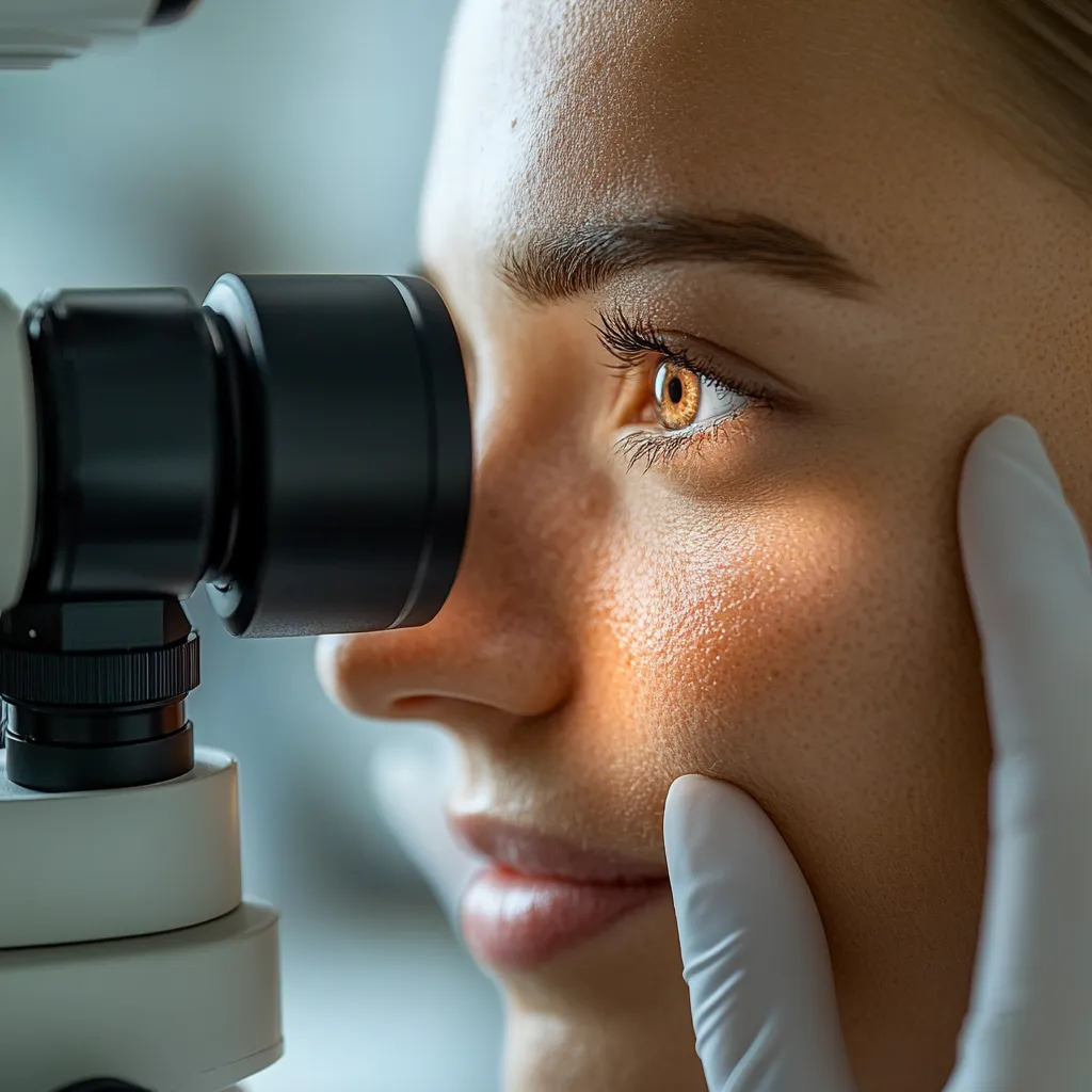
Офтальмология
Международные клинические данные по гирудотерапии при глаукоме, сосудистых заболеваниях сетчатки и воспалительных заболеваниях глаз
Ophthalmologic indications are off-label (Tier B / Tier C). See the Coverage Map for an organ-system view of evidence tiers, and How Evidence Is Graded for methodology.
Исследовательский / Приоритет исследований
Ophthalmic applications of hirudotherapy are not included in FDA 510(k) clearance for medicinal leeches. No ophthalmic indication has received specific regulatory clearance in the United States, the European Union, or any other major jurisdiction.
Исследовательское применение
Международные клинические данные
Ophthalmic hirudotherapy occupies a distinctive position within investigational leech medicine. The eye is an organ of exquisite vascular sensitivity: a rise of a few millimeters of mercury in intraocular pressure, a microvascular occlusion in a retinal arteriole, or a thrombus in the central retinal vein can destroy vision within hours. Published international clinical experience documents the largest cumulative dataset among investigational specialties — over 1,100 patients across glaucoma, retinal vascular disease, inflammatory eye conditions, and visual rehabilitation. The Moscow MNTC “Eye Microsurgery” treated more than 300 patients annually over 15+ years. No randomized controlled trial has been performed for any ophthalmic indication.
Биологическое обоснование
Механизмы глаукомы
Hirudin-mediated anticoagulation may reduce ciliary body secretion; hyaluronidase-facilitated tissue permeability may improve trabecular outflow; histamine-like vasodilation may enhance uveal blood flow. Controlled blood extraction reduces orbital venous pressure.
Сосудистые пути сетчатки
Hirudin inhibits thrombin; destabilase-M lyses cross-linked fibrin; calin and saratin suppress platelet adhesion; lipase/cholesterol esterase fractions exert anti-atherosclerotic effects. Published research describes enhanced retinal perfusion and collateral development.
Противовоспалительный каскад
Eglins block neutrophil elastase and cathepsin G; C1s complement inhibitor modulates classical pathway; hyaluronidase enhances lymphatic drainage; LDTI tryptase inhibitor attenuates mast cell-mediated inflammation in autoimmune ocular conditions.
Клинические доказательства
Уровень доказательности GRADE: Низкий
Наблюдательные исследования или РКИ с серьёзными ограничениями
| Исследование | Дизайн | Популяция (n=) | Вмешательство | Ключевой исход | Результат |
|---|---|---|---|---|---|
| Mozherenkov et al. 1998 | Comparative cohort | Acute angle-closure glaucoma (n=302) | HT (2-3 leeches, temple) + standard tx vs standard tx alone | Attack resolution rate | 87% with HT (n=177) vs 77% without (n=125); p < 0.05 Level III; strongest comparative data for any ophthalmic indication |
| Bondareva & Zhitenev 1998 | Case series | Acute glaucoma — emergency protocol (n=500) | Leeches + acetazolamide + pilocarpine | Pain, congestion, corneal edema | Pain reduction/resolution at 30-40 min post-application Level IV; largest single ophthalmic HT cohort |
| Bagdasarova 1973 | Prospective cohort — tonometric | Chronic glaucoma (n=181) | Leech to temporal region; IOP at 1, 3, 24 h | Intraocular pressure | Significant IOP reduction within 1 h; max at 1-3 h; return toward baseline at 24 h Level IV; consistent temporal pattern |
| Bagdasarova 1973 | Prospective cohort — tonographic | Chronic glaucoma — aqueous dynamics (n=159) | Leech application; tonographic measurement | True IOP, outflow facility, aqueous secretion | All three significantly improved; outflow facility markedly increased Level IV; mechanism data — genuine outflow improvement, not transient volume change |
| Semikova & Bondareva 1995 | Prospective case series | Keratitis, iridocyclitis, uveitis — MNTC program (n=300) | Periorbital application; 6-10 sessions q2-3 days | Infiltrate, tenderness, synechiae, mydriatic response | Corneal clearing; synechiae lysis; enhanced mydriatic effect Level IV; >300 pts/year for >15 years |
| Semikova et al. 1999 | Case series | Post-intraocular surgery complications (n=300) | Single HT session postoperatively | Complication resolution | Single session sufficient in many cases; reduced revision needs Level IV; annual MNTC volume |
| Bondareva 1998 | Case series | Chronic retinal/optic nerve vascular disease (n=12) | 10 sessions; alternating temporal/mastoid; 2-4 leeches | Visual acuity, visual field | VA: 0.01→0.1, 0.1→0.5-0.6, 0.08→0.3, 0.8→1.0; VF +10-20° Level IV; chronic disease typically refractory to standard treatment |
| Mukhanko & Kulanin 2001 | Comparative cohort | Blind/visually impaired — rehabilitation (n=540) | HT + mineral water therapy vs no HT | Visual function improvement | 69% improvement with HT vs 50% controls; no deterioration Level III; combined intervention limits attribution |
| Vedeneeva & Medvedeva 2000 | Case report | Thyroid ophthalmopathy (n=1) | HT for orbital edema, proptosis, diplopia | Clinical response | Successful treatment Level V |
| Yoyleva et al. 1999 | Case series | Optic neuropathies (n=NR) | HT + magnetostimulation | Visual function | Enhanced efficacy with combination therapy Level IV; n not reported |
Глаукома: наиболее убедительные сравнительные данные
Mozherenkov et al. (1998) provided the closest approximation to comparative data: in 302 patients with acute angle-closure glaucoma, the hirudotherapy group achieved 87% attack resolution versus 77% with standard treatment alone (p<0.05). Bondareva and Zhitenev (1998) documented 500 patients receiving leeches alongside acetazolamide and pilocarpine, with pain reduction at 30-40 minutes post-application.
Bagdasarova (1973) conducted tonographic analysis in 340 patients. True IOP decreased, outflow facility increased markedly, and aqueous humor secretion showed compensatory increase — a homeostatic response to improved outflow, not a pathologic rise. These data provide a physiologic explanation for clinical IOP reductions and suggest genuine improvement in outflow physiology rather than transient blood volume reduction.
Данные о физиологическом механизме
Сосудистые заболевания сетчатки: результаты остроты зрения
Bondareva (1998) treated 12 patients with chronic retinal vascular disease (10 sessions, alternating temporal/mastoid). Visual acuity gains were clinically significant: 0.01 to 0.1 (n=2), 0.1 to 0.5-0.6 (n=5), 0.08 to 0.3 (n=3), 0.8 to 1.0 (n=1), with visual field expansion of 10-20 degrees. These represent functional transitions from near-blindness to ambulatory vision in disease stages generally refractory to standard treatment.
Воспалительные заболевания глаз: программа МНТК
Кератит и иридоциклит
Published research documents decreased corneal infiltrate and progressive clearing. Enhanced mydriatic drug action was reported — suggesting SGS-mediated improvement in microcirculatory drug penetration. Posterior synechiae breakdown with pupil dilation documented in iridocyclitis cases.
Увеит и послеоперационный период
Combined therapy produced reduced vitreous precipitates and prevention of coarse vitreous strands. Post-intraocular surgery complications (iritis, macular edema, elevated IOP) managed with single session in many cases, reducing need for additional intervention.
Дополнительные показания
Лучевые осложнения
Semikova (1998) described leech application for radiation-induced inflammation following uveal melanoma treatment. Ocular hemorrhage (hemophthalmos) documented as responsive to SGS fibrinolytic activity.
Оптическая нейропатия
Retrobulbar neuritis and papilledema responded in acute stage. Yoyleva et al. (1999) reported enhanced efficacy when combined with magnetostimulation. Sympathetic ophthalmia particularly emphasized.
Реабилитация
Among 540 patients: 69% improvement with HT vs 50% controls; improvement (n=448) or stabilization (n=92) with no deterioration (Mukhanko & Kulanin 2001).
Клинический протокол — периорбитальное применение
Стандартный протокол
- Preparation: Clean temple/mastoid; no residual antiseptic
- Method: 2 leeches in glass vial pressed against temple or mastoid
- Duration: Until engorgement and spontaneous detachment (20-40 min)
- Course: 6-10 sessions at 2-3 day intervals
- Bilateral: 2 leeches (unilateral) or 4 leeches (bilateral)
Острый приступ глаукомы
- Leeches: 2-3 to temple; +1 mastoid in severe cases
- Assessment: Re-evaluate at 30-40 min for pain/IOP response
- Sessions: Single session may suffice
- Integration: Adjunct to acetazolamide + pilocarpine only
| Показание | Основное место | Альтернатива | Пиявки | Курс |
|---|---|---|---|---|
| Inflammatory disease | Temple (ipsilateral) | Mastoid | 2 | 6-10 sessions, q2-3d |
| Chronic glaucoma | Temple (ipsilateral) | Mastoid | 2 | 6-10 sessions, q2-3d |
| Acute glaucoma | Temple (ipsilateral) | Mastoid (severe) | 2-3 (+1) | Single; reassess 30-40 min |
| Retinal vascular | Temple (alternating) | Mastoid (alternating) | 2 or 4 | 10 sessions |
| Postoperative | Temple (ipsilateral) | Mastoid | 2 | Single session often sufficient |
Предпроцедурное обследование
- Complete ophthalmic exam: VA, IOP (Goldmann tonometry), slit-lamp, dilated fundus
- Visual field testing (automated perimetry) for glaucoma/retinal vascular patients
- CBC, coagulation panel, medication review (anticoagulants, antiplatelets)
- Prior leech exposure and allergy history assessment
- Counseling: periorbital ecchymosis, bleeding duration, cosmetic scarring
Послепроцедурный мониторинг
- Sterile dressing; expect 4 to 24 hours post-detachment oozing
- IOP at 1, 3, and 24 hours (glaucoma patients per Bagdasarova protocol)
- VA and visual field testing at course completion
- Periorbital infection assessment at each session
Вопросы безопасности
Критические требования безопасности
- Globe proximity: Leeches must never be applied to eyelids or conjunctiva. The glass vial containment method is mandatory — prevents migration toward the eye throughout the entire application period.
- Cavernous sinus pathway: The periorbital region drains into the cavernous sinus via valveless ophthalmic veins. Aeromonas hydrophilacould theoretically propagate intracranially. No such case reported; prophylactic antibiotics (fluoroquinolone or TMP-SMX) should be considered in immunocompromised patients.
- Anticoagulant interactions: Patients on warfarin, DOACs, heparin, aspirin, or clopidogrel may experience prolonged bleeding. Coagulation status must be assessed before treatment initiation.
Периорбитальный экхимоз
Post-detachment bleeding may produce “black eye.” Self-limiting, resolving within 7-10 days. Pre-treatment counseling required.
Риск инфекции
Prophylactic antibiotics recommended for immunocompromised patients. Standard wound care. Heightened surveillance for cavernous sinus drainage pathway.
Косметическое рубцевание
Triradiate bite mark heals as small, pale scar. Usually inconspicuous on temple skin. Patients require informed consent regarding facial scarring.
| Препарат | Взаимодействие | Действие |
|---|---|---|
| Warfarin, DOACs, heparin | Additive anticoagulant effect; prolonged bleeding | Assess coagulation; coordinate with prescriber |
| Aspirin, clopidogrel | Additive antiplatelet via calin/saratin | Bleeding time assessment; ecchymosis counseling |
| Mydriatics (atropine, tropicamide) | Enhanced effect reported (improved drug penetration) | Monitor for excessive mydriasis |
| Acetazolamide (oral) | Additive IOP reduction via different mechanisms | Documented in acute glaucoma protocols |
| Prostaglandin analogs | No published data | Theoretical additive benefit; no safety data |
Пробелы в доказательной базе и приоритеты исследований
Методологические ограничения
- No RCTs for any ophthalmic indication
- All evidence Level III-IV (comparative cohorts, case series)
- Multiple combined-intervention studies limit attribution
- Periorbital application inherently difficult to blind
- Geographically homogeneous populations (Russian centers)
- Non-standardized outcome measures across studies
Приоритетные направления исследований
- Pragmatic RCT: adjunctive HT in acute angle-closure glaucoma
- Prospective registry with ETDRS VA, Goldmann IOP, automated VF
- Dose-response: optimal leech count, frequency, course duration
- SGS pharmacokinetics in periorbital tissues
- Long-term follow-up: IOP durability, VA stability at 6-12 months
- Infection surveillance: cavernous sinus pathway risk
Ключевые выводы
Largest investigational evidence base: Over 1,100 patients across glaucoma, retinal vascular, and inflammatory eye disease, with >300 patients/year treated at specialized centers for over 15 years.
Strongest comparative data: 87% vs 77% attack resolution in acute angle-closure glaucoma (p<0.05; Mozherenkov 1998, n=302) — adjunct to standard pharmacologic management.
Documented physiologic mechanism: Tonographic studies (n=340) demonstrate improved aqueous humor outflow facility — genuine physiologic improvement, not transient blood volume reduction.
Multi-target SGS pharmacology: Hirudin (anticoagulation), destabilase (fibrinolysis), hyaluronidase (permeability), eglins (anti-inflammatory), C1s inhibitor (complement), lipase (vascular protection).
Mandatory safety protocol: Glass vial containment prevents globe migration. Cavernous sinus drainage pathway requires heightened infection surveillance. Anticoagulant screening essential.
No RCTs to date: All evidence Level III-IV. Consistent benefit direction across investigators and institutions supports investigation but does not constitute definitive clinical proof.
Список литературы
- Bagdasarova, N. A. (1973). Hirudotherapy in glaucoma: Tonometric and tonographic studies (n=340).
- Bakalova, T. P., Arkhipova, L. T., et al. (2001). Combined therapy for vascular eye disease.
- Bondareva, G. S. (1998). Hirudotherapy in chronic retinal vascular disease (n=12).
- Bondareva, G. S., & Zhiteneva, E. A. (1998). Ophthalmic emergencies and hirudotherapy.
- Chaban, T. N., & Savinov, V. A. (1995). Outpatient hirudotherapy for corneal disease and glaucoma.
- Dorogova, V. B. (1935). Leeches in diseases of the visual organ.
- Ivanova, L. M. (1998). Combined hirudotherapy and acupuncture for retinal vascular disease.
- Kartasheva, E. A. (1979). Leeches in acute angle-closure glaucoma: A critical assessment.
- Mozherenkov, V. P., et al. (1998). Comparative outcomes in acute glaucoma (n=302).
- Mukhanko, L. E., & Kulanin, V. I. (2001). Rehabilitation of visually impaired (n=540).
- Mukhanko, L. E., & Kulanin, V. I. (2003). Extended follow-up of visual rehabilitation.
- Semikova, T. S. (1998). Leech application in choroidal melanoma radiation complications.
- Semikova, T. S., & Bondareva, G. S. (1995). Inflammatory eye disease: MNTC program.
- Semikova, T. S., et al. (1999). Postoperative management with hirudotherapy at MNTC.
- Vedeneeva, Z. V., & Medvedeva, N. G. (2000). Hirudotherapy for thyroid ophthalmopathy.
- Yoyleva, S. A., et al. (1999). Combined HT and magnetostimulation for optic neuropathies.
Связанные ресурсы
Клинические специальности
Все 14 медицинских специальностей.
Гемостаз и коагуляция
Антикоагулянтные механизмы SGS, относящиеся к сосудистым заболеваниям сетчатки.
Ингибиторы протеиназ
Противовоспалительные соединения, относящиеся к воспалению глаз.
Эндокринология
Тиреоидная офтальмопатия — межспециальная связь.
Все показания
Полный список показаний с уровнями доказательности.