Mechanisms of Action
Three Interconnected Pathways of Medicinal Leech Therapy
Mechanism vs. clinical evidence: Of 440+ catalogued salivary proteins, only a fraction underwrites a clinical indication. See the Coverage Map for what is and isn't studied per organ system, the Research Roadmap for ASH's gap priorities, and How Evidence Is Graded.
Educational Content — Mechanism Discussion
Overview: Three Interconnected Pathways
Unlike conventional pharmaceutical interventions that typically target a single receptor, enzyme, or step in a pathological cascade, the application of a living medicinal leech engages at least three distinct but synergistic pathways simultaneously (Baskova, 2004). Understanding these pathways and the manner in which they interact is essential for rational clinical application and for distinguishing evidence-based hirudotherapy from empirical tradition.
1. Local (Humoral) Mechanism
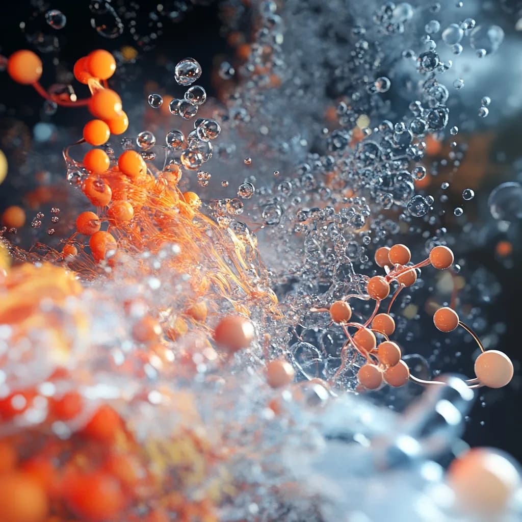
Direct pharmacological action of salivary gland secretion (SGS) components on the wound microenvironment, microcirculatory bed, mast cells, extracellular matrix, and local tissue mediators. Over 100 identified bioactive molecules are delivered directly into the dermal capillary bed.
Onset: Seconds • Strongest evidence base
2. Neuroreflexive Mechanism
Activation of cutaneous sensory afferents by the leech bite, producing segmentally organized somatovisceral reflexes that modulate autonomic nervous system activity and alter the functional state of internal organs sharing the same spinal cord segments.
Onset: Minutes • Explains distant organ effects
3. Systemic (Humoral) Mechanism
Absorption of SGS bioactive compounds into lymphatic and venous circulation, producing measurable changes in coagulation, fibrinolysis, blood rheology, inflammation, and immune function at sites distant from the application point.
Onset: Hours • Critical research gap remains
These three pathways operate on different timescales, creating a temporally layered therapeutic response. Local effects begin within seconds of leech attachment. Neuroreflexive responses develop over minutes as afferent signals reach spinal and supraspinal processing centers. Systemic effects emerge over hours as SGS components distribute through the circulation. This temporal layering may explain why clinical benefits of a single hirudotherapy session often persist for days to weeks — far longer than the pharmacokinetic half-life of any individual SGS component would predict (Baskova, 2004).
Persistent Evidence Gap
Quantitative Session Parameters
| Parameter | Value | Source |
|---|---|---|
| Blood ingested per leech | 5–15 mL | Baskova, 2004 |
| Post-detachment bleeding per wound | Up to 50 mL (30–50 mL typical) | Baskova, 2004 |
| Post-detachment bleeding duration | 4 to 24 hours | Uncontrolled Russian-language report (not independently verifiable) |
| Feeding duration | 20–45 minutes | Baskova, 2004 |
| Wound depth (Y-shaped bite) | ~2 mm | Baskova, 2004 |
| SGS hemostasis blockade duration at wound | 45–60 min (thromboelastographic confirmation) | Uncontrolled Russian-language report (not independently verifiable) |
| Leeches per course (typical) | Up to 50+ | Baskova, 2004 |
| SGS identified bioactive molecules | >100 concurrent (440+ identified in SGS proteome) | Babenko et al., 2020 |
| Anticoagulation factors identified (genomic) | multiple anticoagulation and antihemostatic proteins | Kvist et al., 2020; Babenko et al., 2020 |
Pathway 1: Local (Humoral) Mechanism — SGS Pharmacology
The local mechanism encompasses all direct biochemical and biophysical effects of SGS compounds on the tissue immediately surrounding the bite. Upon biting through the skin, the medicinal leech releases its salivary gland secretion directly into the dermal capillary bed. The SGS contains over 100 identified bioactive molecules (Babenko et al., 2020), each evolved to serve the leech’s feeding requirements but collectively producing a complex pharmacological intervention at the wound site. Genomic and transcriptomic studies have catalogued a large repertoire of salivary proteins across multiple functional categories: analgesic/anti-inflammatory, extracellular matrix degradation, platelet inhibition, anticoagulant, antimicrobial, and others (Babenko et al., 2020).
2.1. The Wound Microenvironment
The leech contributes to local effects through two additional physical mechanisms beyond secretion delivery. First, the three-jawed bite creates a characteristic Y-shaped wound approximately 2 mm in depth, establishing direct access to the superficial dermal vascular plexus. Second, the muscular pharyngeal pump generates negative pressure at the wound site and in adjacent tissues, actively augmenting blood flow to the feeding area (Baskova, 2004). This mechanical suction effect enhances the distribution of SGS components into surrounding tissue and contributes to local decongestive effects.
2.2. Microcirculation Enhancement
The most immediately observable local effect of leech application is the dramatic increase in blood flow to the treatment area. This results from several concurrent processes operating simultaneously:
Vasodilation
Histamine-like compounds and acetylcholine in the SGS produce arteriolar vasodilation in the dermal microcirculation. Capillary perfusion increases markedly, visible as a zone of erythema surrounding the bite site. Measurements using laser Doppler flowmetry have documented significant increases in local blood flow velocity and tissue oxygen saturation during and after leech application (Rothenberger et al., 2016).
Multi-Target Anticoagulation
Hirudin tightly and reversibly binds thrombin with a Kd in the femtomolar range (~20 fM), making it the most potent natural anticoagulant known (Markwardt, 1957). Additional factors — antistasin and lefaxin (factor Xa inhibitors), calin and saratin (platelet adhesion inhibitors), and apyrase (ADP-ase) — collectively block hemostasis at multiple points simultaneously, targeting all three phases of the cell-based coagulation model (Hoffman & Monroe, 2001).
Capillary Permeability Modulation
Hyaluronidase degrades hyaluronic acid in the dermal extracellular matrix, increasing tissue permeability and facilitating the spread of other secretion components through the interstitial space. This "spreading factor" activity amplifies the radius of local pharmacological action well beyond the immediate bite wound. The mechanism has direct parallels in pharmaceutical practice: recombinant hyaluronidase (hylenex) is used clinically to enhance absorption and dispersion of subcutaneously administered drugs.
Net Microcirculatory Effect
The combined result is sustained improvement in local microcirculation: blood flow accelerates, lymphatic drainage improves, venous outflow increases, and tissue edema resolves. Adequate oxygen delivery is restored and metabolic waste products are cleared more efficiently. These effects underlie the well-documented efficacy in microsurgical practice, where leeches salvage congested free flaps and replanted tissues with reported success rates of 65% to 80% (Whitaker et al., 2012).
2.3. Mast Cell Degranulation Modulation
Mast cells, abundant in the dermis, play a central role in the local inflammatory response. SGS components interact with mast cells through multiple receptor-mediated pathways. The leech-derived tryptase inhibitor (LDTI) specifically inhibits mast cell tryptase, a serine protease that promotes inflammation, fibroblast proliferation, and extracellular matrix remodeling (Sommerhoff et al., 1994). By modulating mast cell degranulation rather than simply suppressing it, the SGS creates a controlled local anti-inflammatory environment without eliminating the protective functions of the innate immune response.
Bdellins and eglins — serine protease inhibitors that target neutrophil elastase, cathepsin G, and plasmin — further modulate the inflammatory cascade downstream of mast cell activation. Eglins inhibit the release of proinflammatory cytokines from activated neutrophils, while bdellins reduce the proteolytic tissue damage that accompanies acute inflammation (Baskova & Zavalova, 2001). Together, these inhibitors produce a local anti-inflammatory effect that is functionally distinct from corticosteroids or NSAIDs: rather than broadly suppressing inflammation, they selectively attenuate the tissue-destructive components while preserving the reparative processes.
2.4. Local Anti-Inflammatory Cascade
The anti-inflammatory effect at the local level involves coordinated suppression of multiple inflammatory mediators through functionally distinct pathways:
- Complement modulation — Carboxypeptidase inhibitors in the SGS regulate the complement-derived anaphylatoxins C3a and C5a, reducing chemotaxis of inflammatory cells to the wound site.
- Neutrophil elastase inhibition — Eglins (eglin C in particular) and guamerin inhibit neutrophil elastase with high specificity, preventing the extracellular matrix degradation and tissue edema that characterize acute inflammation.
- Bradykinin regulation — Carboxypeptidase inhibitors also modulate bradykinin metabolism, influencing local vascular permeability and pain signaling.
- Antimicrobial defense — Destabilase, a multifunctional enzyme with both lysozyme (muramidase) and isopeptidase activities, provides antimicrobial protection at the wound site (Kurdyumov et al., 2015). Additional antimicrobial peptides — theromyzin, theromacin, and peptide B — contribute to wound defense against opportunistic pathogens.
Clinically, the local anti-inflammatory effect manifests as reduction of tissue edema, relief of local pain, and acceleration of wound healing. These properties support the established use of hirudotherapy in surgical conditions (thrombophlebitis, varicose veins, postoperative wound complications, abscesses, furuncles), dental practice, trauma management, and inflammatory joint and skin diseases where leeches are applied directly to the affected area (Baskova, 2004).
2.5. Extracellular Matrix Remodeling
Hyaluronidase occupies a unique position among SGS components. By catalyzing the hydrolysis of hyaluronic acid — a major structural component of the dermal extracellular matrix — it increases tissue permeability and facilitates the interstitial diffusion of all other SGS compounds. Beyond its spreading function, hyaluronidase-mediated matrix remodeling may contribute to the resolution of fibrotic tissue changes associated with chronic inflammation. The degradation of hyaluronic acid generates oligosaccharide fragments that themselves possess immunomodulatory properties, potentially contributing to the anti-inflammatory milieu at the application site.
The tissue penetration facilitated by hyaluronidase is pharmacologically significant for OA applications. Experimental data on topical diclofenac gel applied to knees with joint effusions demonstrated drug detection in deep periarticular tissues and body compartments. With the additive effect of hyaluronidase as a spreading factor, it is highly probable that the antiphlogistic substances in leech SGS penetrate deep enough to exert significant effects on periarticular myofascial structures and perhaps even on intra-articular structures (Michalsen et al. 2007). A recent study confirmed that periarticular myofascial structures play a critical role in the development of chronic joint pain and regional pain syndromes in patients with osteoarthritis.
Hyaluronidase activity (a 'spreading factor') in leech SGS facilitates diffusion of salivary components through host extracellular matrix. This activity suggests that the leech has evolved a coordinated approach to tissue penetration that simultaneously serves its feeding requirements and — incidentally — produces therapeutic matrix-remodeling effects in the host tissue.
2.6. Complete SGS Compound Categories at the Local Level
| Category | Compounds | Molecular Targets | Local Effect | Duration |
|---|---|---|---|---|
| Anticoagulants | Hirudin, antistasin, lefaxin, ghilanten | Thrombin (Kd ~20 fM), factor Xa (Ki ~0.5 nM), factor XIIIa (transglutaminase) | Complete hemostasis blockade at the bite wound; multi-target inhibition across all three phases of cell-based coagulation model | 4 to 24 hours post-detachment bleeding |
| Platelet inhibitors | Calin, saratin, decorsin, apyrase, PAF inhibitor | Collagen–platelet adhesion, vWF–collagen interaction (shear-dependent), GP IIb/IIIa integrin, extracellular ADP, platelet-activating factor | Prevention of platelet plug formation at the wound site; blockade of primary hemostasis | Sustained via collagen-bound calin (irreversible binding) |
| Vasodilators | Histamine-like compounds, acetylcholine | Arteriolar smooth muscle, endothelial NO release | Arteriolar vasodilation in dermal microcirculation; increased capillary perfusion; visible erythema zone surrounding bite | Minutes to hours |
| Anti-inflammatory agents | Bdellins, eglins (eglin C), LDTI, guamerin, carboxypeptidase inhibitors | Neutrophil elastase, cathepsin G, plasmin, mast cell tryptase, complement C3a/C5a, bradykinin metabolism | Selective attenuation of tissue-destructive inflammation while preserving reparative processes; functionally distinct from corticosteroids or NSAIDs | Hours; sustained by continued SGS presence in wound |
| ECM remodeling enzymes | Hyaluronidase, collagenase | Hyaluronic acid (major ECM structural component), collagen fibers | 'Spreading factor' activity: increases tissue permeability and facilitates interstitial diffusion of all other SGS compounds; radius of pharmacological action extends beyond immediate bite wound | Hours; analogous to pharmaceutical hylenex (recombinant hyaluronidase) |
| Antimicrobial agents | Destabilase (lysozyme + isopeptidase), theromyzin, theromacin, peptide B | Bacterial peptidoglycan (muramidase activity), bacterial cell membranes | Wound defense against opportunistic pathogens; dual enzymatic and non-enzymatic antimicrobial mechanisms | Sustained during wound healing period |
2.7. Evidence Base for Local Mechanisms
The local mechanism is supported by the strongest evidence among the three pathways:
- In vitro studies — Individual SGS components have been characterized biochemically and their targets identified at the molecular level. Draft-genome and salivary-transcriptome studies of H. medicinalis (Babenko et al., 2020) have identified multiple anticoagulant and antihemostatic salivary proteins, including new homologs of genes encoding known anticoagulants, across several functional categories.
- Animal models — Leech application produces measurable changes in local blood flow, tissue oxygenation, and wound healing parameters in standardized animal models.
- Clinical observation — Microsurgical flap salvage represents the highest level of clinical evidence for local mechanisms, with a systematic review documenting an overall success rate of ~78% (216 of 277 cases) (Whitaker et al., 2012). Tissue spectrophotometry and laser Doppler flowmetry studies have quantified the magnitude and duration of microcirculatory changes (Rothenberger et al., 2016).
- Pharmacological validation — Multiple SGS-derived compounds have progressed to pharmaceutical development, including three FDA-approved direct thrombin inhibitors (lepirudin, bivalirudin, desirudin) and recombinant destabilase in preclinical development for aged thrombus dissolution (Kurdyumov et al., 2015).
From Leech to Pharmacy
Pathway 2: Neuroreflexive Mechanism — Dermatomal Somatovisceral Reflexes
If hirudotherapy were purely a local pharmacological intervention, the site of leech application would matter only insofar as it determines which tissues receive the direct effects of SGS. Traditional hirudotherapy practice holds that the site of application can influence outcomes in distant organs (Baskova, 2004) — observations that are hypothesized to involve reflex mechanisms but have not been confirmed in controlled studies.
3.1. Dermatomal Organization: The Head Framework
The neuroanatomical foundation for the reflexive pathway rests on the principle of dermatomal organization — the segmental arrangement of sensory innervation established during embryonic development. Each spinal nerve innervates a defined strip of skin (a dermatome) and, through its visceral branches, a corresponding set of internal organs. This dual innervation creates bidirectional neural pathways between the body surface and the viscera.
Grigory Zakharyin (1889) described that diseases of internal organs produce referred pain and altered sensitivity in specific cutaneous areas. He observed that during or following an anginal episode, touching the patient’s skin at certain body areas produced pain. Henry Head (1898) subsequently mapped these zones of cutaneous hyperalgesia systematically, establishing the association between visceral disease and altered skin sensitivity in corresponding dermatomes.
The Head zones are now understood through the lens of modern neuroanatomy as a manifestation of viscerosomatic convergence — the convergence of visceral and somatic afferent neurons at common spinal cord segments. Smolin (1982) and Cervero & Tattersall (1985) demonstrated that visceral and cutaneous afferent fibers converge onto the same dorsal horn neurons. Because the cerebral cortex receives far more somatosensory than visceral input, it misinterprets visceral nociceptive signals arriving through shared spinal pathways as originating from the corresponding dermatome. This "convergence-projection" mechanism explains referred pain and, critically, also explains why stimulation of the appropriate dermatome can modulate visceral function.
The convergence operates at multiple levels of the neuraxis. Cervero & Connell (1983) showed that visceral-cutaneous signal interaction begins at the level of primary sensory neurons in the dorsal root ganglia. The interaction extends through the dorsal horn of the spinal cord, where somatic afferents may synapse onto visceroreceptor neurons in the lateral horns, and continues through the brainstem (reticular formation), thalamus, and cortex (Nozdrachev, 1983). The extensive vertical integration of this convergence ensures that cutaneous stimulation can produce both spinal and supraspinal effects on visceral function.
3.2. Segmental Innervation of Major Organs
The following table summarizes the segmental innervation of major internal organs, providing the neuroanatomical basis for application site selection in hirudotherapy (adapted from Table 19, Baskova, 2004):
| Organ | Sympathetic Segments | Parasympathetic Innervation |
|---|---|---|
| Heart | T1–T5 | Vagus nerve, phrenic nerve (C2–C5) |
| Lungs and bronchi | T1–T5 | Vagus nerve, phrenic nerve (C2–C5) |
| Stomach (cardia) | T5–T7 | Vagus nerve, phrenic nerve (C2–C5) |
| Stomach (body) | T7–T8 | Vagus nerve, phrenic nerve |
| Stomach (pylorus) | T8–T9 | Vagus nerve |
| Small intestine, ascending colon | T9–L1 | Vagus nerve |
| Descending colon, rectum | L1–L3 | S2–S5 |
| Liver, gallbladder | T7–T11 | Vagus nerve, phrenic nerve (C2–C5) |
| Pancreas | T8 | Vagus nerve |
| Kidneys and ureter | T8–L2 | — |
| Urinary bladder (body) | T11–L2 | S2–S4 |
| Prostate gland | T10–T12 | S1–S2 |
| Uterus | T12–L1 | S2–S4 |
Clinical significance: The heart receives sympathetic innervation from segments T1 through T5 and parasympathetic innervation from C2 through C5. The Head zones for cardiac disease therefore span dermatomes C3 through T5 — precisely the cutaneous areas where patients with angina pectoris report referred pain along the medial surface of the arm and forearm, in the precordial region, and in the interscapular area. Head documented that in angina pectoris, generalized zones of hyperalgesia appeared in dermatomes C3–C4 and T1–T5, sometimes extending bilaterally (Head, 1898).
3.3. Objective Detection of Dermatomal Changes: Biophysical Methods
The viscerocutaneous reflex produces not only subjective hyperalgesia but also objectively measurable changes in the biophysical properties of the skin within the corresponding dermatomes. These include alterations in vasomotor activity, sweat gland function, skin temperature, electrical potential and capacitance, and resistance to electric current (Borodulin, 1963; Voll, 1973; Warren, 1976; Eory, 1984).
Pain Sensitivity Testing
Bykhovskaya & Eydinova (1935) developed a method for detecting hyperalgesia zones based on adaptation time to pinprick stimulation. In 20 healthy subjects, bilateral adaptation occurred within 3 to 7 seconds. In patients with angina, Levinson (1948) documented prolongation to 40 to 60 seconds, with occasional intensification and spatial spread of the pain sensation.
Cutaneous Thermography
Schneider-Bibus (1986) measured thoracic surface temperatures in 28 patients with MI, CAD, and arrhythmias, developing a schema for localizing points of reduced temperature corresponding to cardiac dermatomes. Borodulin (1963) established that thermoasymmetry of 0.5 to 1.0 °C between symmetric points was normal, while values exceeding this threshold were pathological. Schneider-Bibus demonstrated clear correlation between thermographic data and ECG, angiographic, and myocardial scintigraphy findings.
This claim cites a study that could not be located in any bibliographic database and should be removed; no verifiable source supports the reported patient counts or measurements.
These biophysical methods confirm that Head zones correspond to objectively measurable physiological alterations in the dermatomal skin, consistent with contemporary understanding of central sensitization: visceral nociceptive input dynamically alters the excitability of dorsal horn neurons, producing transient and spatially variable changes in cutaneous sensitivity within the corresponding dermatomes (Woolf, 2011).
3.4. Gate Control Theory and Pain Modulation
The gate control theory of pain, proposed by Melzack & Wall in their landmark 1965 paper in Science, provides a foundational framework for understanding how the leech bite — a peripheral cutaneous stimulus — can modulate pain perception. The theory proposes that a neural mechanism in the dorsal horn acts as a "gate" modulating nociceptive transmission:
- Large-diameter myelinated A-beta fibers (conveying touch, pressure, vibration): activity excites inhibitory interneurons in the substantia gelatinosa (lamina II), which suppress second-order transmission neurons — effectively "closing the gate".
- Small-diameter C fibers and A-delta fibers (conveying nociceptive signals): activity inhibits the inhibitory interneurons — "opening the gate" and facilitating pain transmission.
When the leech bites, it simultaneously activates both mechanoreceptors (via mechanical pressure of the three-jawed bite) and nociceptors (via tissue damage). The sustained mechanical stimulation of A-beta fibers during feeding — which lasts 20 to 45 minutes — activates the gate control mechanism, reducing nociceptive transmission from the same and adjacent dermatomes. This is the same principle exploited by transcutaneous electrical nerve stimulation (TENS) and the intuitive human behavior of rubbing an injured area (Sluka & Walsh, 2003).
However, gate control theory alone cannot fully account for the analgesic effects of hirudotherapy. The theory predicts analgesia lasting only as long as the large-fiber stimulation continues. Clinical pain relief from leech therapy persists for hours to days. Additional mechanisms must therefore be involved.
3.5. Descending Pain Modulation: PAG-RVM Pathway
Beyond the spinal gate, the CNS has a powerful descending pain modulatory system originating in the brainstem and projecting to the spinal cord dorsal horn. Two principal structures are involved:
Periaqueductal Gray (PAG)
Located in the midbrain surrounding the cerebral aqueduct. Receives convergent input from cortex, hypothalamus, amygdala, and ascending spinal pathways. The ventrolateral PAG (vlPAG) sends excitatory projections to the RVM, activating descending inhibition of spinal nociceptive transmission (Heinricher et al., 2009).
Rostroventromedial Medulla (RVM)
Contains functionally distinct "on-cells" (facilitate pain) and "off-cells" (inhibit pain). Activation of off-cells produces spinal analgesia through serotonergic, noradrenergic, and opioidergic mechanisms. Also projects to the locus coeruleus, engaging noradrenergic descending inhibition (Luskin et al., 2023).
3.6. Conditioned Pain Modulation (CPM / DNIC)
Peripheral noxious stimulation — including the sustained nociceptive input from a leech bite — can activate the descending modulatory system through the phenomenon known as diffuse noxious inhibitory controls (DNIC), now more commonly termed conditioned pain modulation (CPM). First described by Le Bars et al. (1979), DNIC represents a "pain-inhibits-pain" mechanism in which noxious stimulation at one body site inhibits second-order wide dynamic range (WDR) neurons at distant spinal segments.
The neurochemical mediators of DNIC include norepinephrine (acting through spinal alpha-2 adrenoreceptors), serotonin (acting through 5-HT receptors in the dorsal horn), and endogenous opioids (acting through mu and delta receptors in the PAG and RVM). The involvement of multiple neurotransmitter systems suggests that activation by the leech bite produces a strong and sustained antinociceptive response (Yarnitsky, 2010).
This mechanism provides a neurophysiological explanation for the observation that leech application at one body site can reduce pain perception at distant sites. The sustained nociceptive input from the bite wound — which continues for hours during post-detachment bleeding — may produce prolonged activation of the descending inhibitory system.
3.7. Additional Analgesic Mechanisms
Local Adenosine Release
Goldman et al. (2010) demonstrated in Nature Neuroscience that peripheral tissue stimulation triggers adenosine release acting on A1 receptors to produce local anti-nociception persisting beyond the stimulus. The mechanical tissue disruption and inflammatory milieu from the leech bite would promote adenosine release from damaged cells and activated immune cells.
Neuroplastic Cortical Changes
Repeated peripheral stimulation produces measurable changes in primary somatosensory cortex (S1) organization. Neuroimaging meta-analyses document altered functional connectivity in bilateral frontal gyrus, temporal gyrus, inferior parietal lobe, parahippocampus, and precuneus (Zhang et al., 2025). These adaptations may explain progressive improvement over serial hirudotherapy courses.
3.8. Integrated Temporal Model of Analgesia
The multiple analgesic mechanisms can be organized into a temporal sequence that explains the characteristic clinical pattern of pain relief following hirudotherapy:
| Phase | Timescale | Mechanism | Description | Reference |
|---|---|---|---|---|
| Immediate | Seconds | Gate control | Mechanical stimulation of large-diameter A-beta fibers closes the spinal gate to nociceptive transmission in the dorsal horn substantia gelatinosa (lamina II) | Melzack & Wall, 1965 |
| Short-term | Minutes | Bioactive saliva + adenosine | Anti-inflammatory eglins and bdellins reduce nociceptor sensitization; poorly characterized anesthetic compounds; local adenosine release via A1 receptors provides sustained anti-nociception independent of neural transmission | Goldman et al., 2010 |
| Medium-term | Minutes to hours | CPM / descending modulation | Somatoautonomic reflexes modulate visceral function and autonomic tone. Conditioned pain modulation (DNIC) provides supraspinal descending inhibition via PAG–RVM pathway through noradrenergic, serotonergic, and opioidergic mechanisms | Le Bars et al., 1979; Yarnitsky, 2010 |
| Long-term | Days to weeks | Neuroplastic adaptation | Sustained anti-inflammatory effects from continued wound healing and decongestive bleeding; improved local microcirculation; neuroplastic changes in cortical pain processing over repeated sessions (bilateral frontal gyrus, temporal gyrus, inferior parietal lobe, parahippocampus, precuneus) | Zhang et al., 2025 |
This multi-layered temporal model explains why clinical pain relief from hirudotherapy often exceeds what would be predicted by any single mechanism in isolation.
3.9. Somatoautonomic Reflexes — The Core Therapeutic Mechanism
The most therapeutically significant aspect of the neuroreflexive pathway is not pain modulation per se, but the activation of somatoautonomic reflexes — neural pathways through which cutaneous stimulation modulates autonomic nervous system activity and, thereby, visceral organ function. Systematically characterized by Sato & Schmidt beginning in 1966, the somatoautonomic reflex follows a defined arc (Sato et al., 1997; Li et al., 2016):
- Stimulation of cutaneous mechanoreceptors and nociceptors activates somatic afferent neurons (cell bodies in dorsal root ganglia)
- Afferent signals transmitted to spinal cord, processed and relayed to autonomic efferent neurons
- Postganglionic fibers terminate on adrenergic or cholinergic effector cells in target organs
- Sympathetic or parasympathetic responses produced in target organ
The critical feature is segmental specificity. Cutaneous stimulation at a given dermatomal level preferentially activates autonomic pathways innervating organs sharing the same spinal cord segments. Somatic afferents entering the spinal cord switch onto visceroreceptor neurons in the lateral horns (intermediolateral column, T1–L2), giving rise to preganglionic sympathetic fibers. This means stimulation of dermatomes T1–T5 preferentially modulates cardiac autonomic function, T7–T11 preferentially modulates hepatobiliary function, and so on (Sudakov et al., 1986).
The postganglionic fibers terminate on both adrenergic and cholinergic neurons, meaning cutaneous dermatomal stimulation can produce either sympathetic or parasympathetic effects: vasospasm or vasodilation, slowing or acceleration of intestinal peristalsis, modulation of glandular secretion (Sudakov et al., 1986). The direction and magnitude depend on stimulus intensity, duration, modality, and the baseline autonomic tone of the target organ.
Liu et al. (2020) demonstrated two key organizational principles directly relevant to hirudotherapy practice:
- Acupoint selectivity: specific skin locations activate distinct autonomic pathways, validating the practice of selecting application sites based on their segmental relationship to the target organ.
- Intensity dependence: the strength of the cutaneous stimulus determines which autonomic pathways are activated. The leech bite, producing sustained moderate-intensity nociceptive and mechanical stimulation over 20–45 minutes of feeding followed by hours of wound bleeding, represents a stimulus profile well suited to sustained autonomic modulation.
Modern Neuroimaging Confirmation
3.10. The Cutaneous-Visceral Reflex in Practice: Clinical Demonstration
When leeches are applied to the precordial region for angina pectoris, the bite stimulates cutaneous afferents in dermatomes T1 through T5 — the same spinal segments providing sympathetic innervation to the heart, so cutaneous stimulation of this zone can plausibly engage cardiac sympathetic reflex pathways.
Warming the Left Hemithorax
A cutaneous–visceral reflex between the skin and the coronary circulation — whereby dilation of cutaneous vessels is accompanied by dilation of coronary vessels — has been described only in uncontrolled Russian-language reports that are not independently verifiable. No quantified or peer-reviewed data are available to support this mechanism.
Chloroethyl Spray (Cooling)
Produced a biphasic response: initial cutaneous cooling with worsening myocardial ischemia and pain intensification, followed by a more prolonged phase of coronary vasodilation and pain relief as the local vascular response reversed. Constriction of cutaneous vessels was accompanied by corresponding constriction of coronary vessels.
These observations establish that the clinical zones traditionally used in hirudotherapy are designated application zones — extensive cutaneous areas within the dermatomes that share segmental innervation with the target organ. The therapeutic stimulus from any point within this dermatomal zone is mediated via the somatovisceral reflex pathway. This is the same principle that underlies all forms of cutaneous reflex therapy: TENS, thermotherapy, mustard plasters, topical counter-irritants, and procaine blocks (Baskova, 2004).
3.11. Comparison with Other Cutaneous Therapies
Hirudotherapy is not the only therapeutic modality that exploits cutaneous-visceral reflexes, but it is unique in combining neuroreflexive stimulation with simultaneous pharmacological delivery. The following comparison clarifies its distinctive features:
| Modality | Stimulus Type | Fiber Activation | Mechanism | Pharmacological? |
|---|---|---|---|---|
| TENS (conventional) | Low-intensity, high-frequency electrical | A-beta only (selective) | Gate control (spinal gating) | None |
| TENS (acupuncture-like / intense) | High-intensity, low-frequency electrical | A-beta + A-delta/C fibers | Spinal gating + descending modulation | None |
| Acupuncture | Metal needle, point-based mechanical | A-beta + A-delta + C fibers | Somatoautonomic reflex via segmental innervation; identical dermatomal principle | None |
| Hirudotherapy | Y-shaped bite + pharyngeal suction (20–45 min feeding + hours of wound bleeding) | A-beta + A-delta + C fibers (simultaneous mechanoreceptor + nociceptor activation) | Gate control + descending modulation + somatoautonomic reflexes | 100+ bioactive SGS compounds delivered concurrently |
| Thermotherapy (heating pads, cold packs) | Thermal (warming or cooling) | Thermoreceptors + nociceptors | Cutaneous–visceral reflex; warming precordial area improves coronary perfusion | None |
| Mustard plasters / capsaicin | Chemical irritant (topical counter-irritation) | C fibers (TRPV1 activation) | Counter-irritation; dermatomal reflex; conditioned pain modulation | None (surface-acting only) |
Neurophysiological research has demonstrated that acupuncture effects are mediated by the nervous system — specifically, by cutaneous-visceral reflexes operating through segmental innervation pathways (Peng, 1986). The meridians defined in traditional Chinese medicine correspond anatomically to the topography of peripheral nerve trunks and neurovascular bundles (Dung, 1984). The designated application sites used in acupuncture are located within the same dermatomes as the broader Head zones — the coincidence is not accidental, because both reflect the underlying segmental innervation (Pishel et al., 1995).
The key distinction between acupuncture and hirudotherapy as reflex therapy methods lies in stimulus characteristics: acupuncture delivers a point-based mechanical stimulus from a metal needle at precise anatomical locations, while hirudotherapy delivers a broader stimulus from a biologically active zone. A course of hirudotherapy typically requires up to 50 or more leeches; the cutaneous zones must be territorially sufficient to accommodate this number, a requirement better served by dermatomal zones than by individual acupuncture points.
3.12. Application Site Selection: Dermatomal Rationale
The neuroanatomical evidence establishes that the traditional practice of applying leeches to specific body regions is not an empirical survival from pre-scientific medicine but a rational therapeutic strategy grounded in the segmental organization of the nervous system. Application site selection follows a clear algorithm:
- Identify the target organ and its spinal segmental innervation (see segmental innervation table above).
- Map the corresponding dermatomes on the body surface.
- Apply leeches within these dermatomes, selecting areas that are accessible, well-vascularized, and distant from major vessels and nerves.
This reframing has practical significance: relocating leeches within the appropriate dermatomal zone does not fundamentally alter the reflexive mechanism of action. Whether leeches are placed at specific acupuncture points or distributed across the broader reflex zone, the afferent signals reach the same spinal cord segments and activate the same somatoautonomic reflex arc. The apparent coincidence between traditional hirudotherapy zones, Head zones, and acupuncture meridian distributions reflects the same underlying dermatomal neuroanatomy (Baskova, 2004).
Pathway 3: Systemic (Humoral) Mechanism — Circulating SGS Compounds
The third pathway of therapeutic action involves the entry of SGS bioactive compounds into the lymphatic and venous circulation, where they produce effects at sites distant from the application point. Two pathways of systemic distribution have been proposed (Baskova, 2004):
Intercellular Signaling
SGS components act on receptors of various cell types through signal transduction mechanisms. Baskova and colleagues demonstrated that SGS influences the transport of monovalent cations and calcium ions in human platelets through receptor-mediated pathways. These signaling effects can propagate through local cell populations and, through circulating cellular elements (platelets, leukocytes), extend to distant sites.
Direct Vascular Absorption
Any pharmacological agent delivered intradermally or subcutaneously will inevitably enter the vascular bed. The leech releases SGS directly into the dermal capillary bed, where blood flow velocity is low and intravascular pressure is modest. While some SGS enters the leech’s gut with ingested blood, and a portion is consumed in local hemostatic interactions, a significant fraction remains in the wound and surrounding tissues available for absorption.
4.1. Evidence of Prolonged Local SGS Activity
The prolonged local bleeding that follows leech detachment — typically lasting 24 to 48 hours — is consistent with anticoagulant components of the salivary gland secretion remaining bioactive in the wound for an extended period. The longer these components remain in the wound area, the greater the opportunity for absorption into blood and lymphatic circulation.
The lymphatic system plays a particularly important role. Lymphatic microvessels draining the dermis continuously absorb interstitial fluid and return it to the venous circulation. Under normal physiological conditions, 60% of plasma volume and 45% of plasma proteins transit from the microcirculation into the interstitial space daily, carrying protein-bound metabolites into the lymphatic system before returning to the bloodstream (Chernukh & Frolov, 1982). SGS components deposited in the dermal interstitium would be subject to this same lymphatic clearance mechanism. Hyaluronidase — by increasing tissue permeability through ECM degradation — facilitates the interstitial diffusion and subsequent lymphatic absorption of other SGS compounds.
4.2. Hirudin Pharmacokinetics (Reference Data)
The pharmacokinetics of recombinant hirudin have been studied extensively in the context of drug development, providing a framework for understanding the systemic anticoagulant effects. These parameters, derived from purified recombinant protein, provide only an approximation of native hirudin behavior released into a bite wound:
| Parameter | IV Administration | SC Administration | Source |
|---|---|---|---|
| Distribution half-life (t½α) | 5–15 min | N/A | Markwardt et al., 1984 |
| Elimination half-life (t½) | ~60 min | ~120 min | Markwardt et al., 1984 |
| Volume of distribution | ~12.9 L | ~12.9 L | Bichler et al., 1991 |
| Bioavailability | 100% (reference) | ~100% | Markwardt et al., 1984 |
| Elimination route | ~90% renal | ~90% renal | Bichler et al., 1991 |
| PK model | One-compartment | One-compartment | Markwardt et al., 1984 |
4.3. Systemic Anticoagulant Effects
Clinical observations support the occurrence of systemic anticoagulant effects. Baskova (2004) reported that the anticoagulant effect was observed regardless of the site of leech application — precordial area, right hypochondrium, or mastoid process — suggesting a systemic rather than purely local mechanism. Platelet aggregation inhibition has also been documented as a systemic effect, independent of cutaneous application site, potentially mediated by calin, saratin, and decorsin.
A clinical observation illustrates the phenomenon: a patient experienced profuse bleeding for 12 hours after leech detachment, prompting a surgeon to apply metal staples to the wounds; blood then seeped from the staple wounds as well, and hemostasis was ultimately achieved only with a tight pressure dressing applied over the entire operative field (Baskova, 2004).
4.4. Systemic Anti-Inflammatory Effects
Beyond anticoagulation, several SGS components have the potential to produce systemic anti-inflammatory effects upon entering the circulation:
- Complement modulation — SGS inhibitors of complement-derived anaphylatoxins and carboxypeptidases could modulate the complement cascade at distant sites, consistent with the convergent model of coagulation recognizing that coagulation and innate immune activation are interconnected (Yong & Toh, 2023).
- Cytokine suppression — Bdellins and eglins, through neutrophil serine protease inhibition, reduce release of proinflammatory cytokines (IL-1β, IL-6, TNF-α) from activated neutrophils and macrophages. At sufficient systemic concentrations, these could attenuate the systemic inflammatory response.
- Immune modulation — Clinical observations have documented changes in immune parameters: modulation of lymphocyte subpopulations and immunoglobulin levels. The presence of lectins, ficolins, and CRISP proteins in the secretion (Babenko et al., 2020) suggests multiple pathways of immune interaction.
Experimental Validation: Lausanne PEG Hirudin Study
4.5. Blood Rheology Changes
Hirudotherapy produces measurable changes in blood rheological parameters documented across multiple clinical studies:
- Decreased blood viscosity — Attributed to anticoagulant and fibrinolytic effects of absorbed SGS, as well as mechanical depletion of blood volume through ingestion (5–15 mL per leech) and post-detachment bleeding (50–150 mL per wound site).
- Reduced erythrocyte aggregation — Potentially mediated by calin and other platelet/cell adhesion inhibitors.
- Improved erythrocyte deformability — Possibly resulting from reduced fibrinogen levels and altered plasma protein composition.
These rheological changes improve microcirculatory flow characteristics and may contribute to clinical benefits in conditions characterized by hyperviscosity: chronic venous insufficiency, polycythemia, and microvascular complications of diabetes.
In a cohort of 23 patients receiving a single leech treatment in the lumbar region, reduced viscoelasticity and aggregation tendency of the blood were observed four weeks after treatment, while hematocrit and plasma viscosity values remained unchanged. Given the short plasma half-life of hirudin (~60 minutes IV, ~120 minutes SC), the authors proposed that differential stimulation of erythropoiesis might be responsible for this long-term modulation of hemorheological parameters (Michalsen et al. 2007, citing Kalender et al.).
4.6. Lipid Metabolism Effects — An Intriguing Observation
Since lipid metabolism is primarily regulated by the liver, and the liver’s reflex zone encompasses dermatomes T7–T11, any lipid-related effect observed with precordial (T1–T5) or mastoid application could not be attributed to a local reflexive pathway alone.
This observation suggests that either the systemic mechanism contributes through absorbed SGS components acting on hepatic or systemic metabolic pathways, or that the reflexive mechanism operates through more complex intersegmental pathways than simple dermatomal correspondence would predict. The question remains open and warrants investigation with modern metabolomics and pharmacokinetic methods.
4.7. Evidence Base for Systemic Mechanisms
Evidence Limitation
- Pharmacological reasoning — All intradermally or subcutaneously administered substances enter the systemic circulation. Recombinant hirudin has nearly 100% bioavailability after subcutaneous injection (Markwardt et al., 1984).
- Clinical observation — Anticoagulant and antiplatelet effects observed regardless of application site, implying systemic distribution.
- Blood marker studies — Multiple investigators have documented changes in coagulation parameters, platelet aggregation, blood viscosity, and lipid profiles following hirudotherapy.
- Temporal profile — Some therapeutic effects emerge over hours and persist for days, consistent with the pharmacokinetic time course of absorbed and distributed SGS components.
A critical research priority is the direct measurement of SGS components (particularly hirudin, destabilase, and eglins) in the blood of patients undergoing hirudotherapy, using modern proteomic or immunoassay methods. Such studies would definitively establish the systemic pathway and provide the pharmacokinetic basis for rational dosing.
4.8. Negative Correlations: What Does Not Predict Outcome
Two large studies on leech therapy in knee OA provided sub-analyses that tested whether certain baseline parameters predicted treatment response (Michalsen et al. 2007). The results were consistently negative:
- Connective tissue zones: No correlation between the extent of local connective tissue zones (Bindegewebszonen) and clinical efficacy. Some practitioners postulate that leech therapy is especially effective in patients with pronounced connective tissue changes, but this was not supported by the data.
- Hematocrit and blood volume: No correlation between initial hematocrit levels, the volume of blood extracted by the leech, or BMI (used as approximate constitutional parameters) and treatment outcome.
- Lymphedema: Clinical efficacy was demonstrated even in patients without palpable tissue abnormalities or lymphedema, suggesting that stimulation of lymph flow, while potentially relevant in varicosis, is of limited relevance to the pain-relieving mechanism.
These negative findings are clinically significant: they indicate that the multi-target mechanism of hirudotherapy operates independently of the patient’s constitutional type or humoral parameters. The Pischinger basic regulation model, which focuses on connective tissue protein storage and ground substance regulation rather than cellular processes, provides a theoretical framework for this observation, though its relevance to the pain-relieving effect remains speculative.
Integration: How the Three Mechanisms Synergize
The three mechanisms of hirudotherapy do not merely coexist — they interact synergistically in ways that may amplify the therapeutic effect beyond what any single mechanism would achieve alone. Understanding these interactions is critical for rational clinical application.
5.1. Temporal Sequence of the Three Pathways
| Timescale | Pathway | Key Events |
|---|---|---|
| Seconds to minutes | Local | SGS release into wound. Local anticoagulation, vasodilation, and mast cell modulation commence within seconds. Pharyngeal pump generates negative pressure augmenting blood flow. Within minutes: local anti-inflammatory cascade fully engaged, ECM remodeled by hyaluronidase, antimicrobial defense established. |
| Minutes to tens of minutes | Neuroreflexive | Afferent signals reach spinal cord within milliseconds, but full somatoautonomic reflex responses require minutes of sustained input. Gate control analgesia begins immediately. Descending pain modulation (PAG-RVM) engaged by nociceptive component. Conditioned pain modulation develops. Autonomic modulation builds over the 20–45 min feeding period and persists as post-detachment bleeding maintains afferent input. |
| Hours to days | Systemic | Gradual absorption of SGS components into lymphatic and venous circulation. Systemic anticoagulant, anti-inflammatory, and rheological effects develop. Depletion of blood volume through ingestion (5–15 mL per leech) and post-detachment bleeding (50–150 mL per wound site) produces hemodilution improving blood rheology. |
5.2. Synergistic Interactions Between Pathways
Local + Reflexive
Forward Enhancement
SGS-mediated vasodilation enhances dermal blood flow, increasing the density of activated sensory receptors per unit area and amplifying the cutaneous–visceral reflex signal
Reverse Enhancement
Autonomic reflex response enhances target organ blood flow, improving the distribution of locally delivered SGS to deeper tissue layers
Local + Systemic
Forward Enhancement
Hyaluronidase-mediated increase in tissue permeability facilitates absorption of SGS components into lymphatic and venous circulation, amplifying the systemic mechanism
Reverse Enhancement
Prolonged local bleeding maintained by local anticoagulation provides a sustained source of SGS for systemic absorption and a continuous nociceptive stimulus that maintains reflex activation
Reflexive + Systemic
Forward Enhancement
Autonomic modulation of target organ blood flow (via somatovisceral reflex) enhances delivery of systemically absorbed SGS compounds to the target tissue
Reverse Enhancement
Systemic anti-inflammatory effects of distributed eglins and bdellins reduce peripheral nociceptive input from the underlying pathology, potentiating descending pain modulation
5.3. Why Hirudotherapy May Exceed the Sum of Its Parts
The multi-mechanism nature of hirudotherapy distinguishes it from single-mechanism pharmaceutical interventions. A conventional anticoagulant (e.g., warfarin, heparin, or a DOAC) targets one step in the coagulation cascade. Hirudotherapy simultaneously engages local anticoagulation at multiple cascade steps, neuroreflexive modulation of organ function, and systemic anti-inflammatory and rheological effects.
The analogy to combination pharmacotherapy is instructive. In cardiovascular medicine, combination regimens (anticoagulant plus antiplatelet plus antihypertensive plus lipid-lowering agent) are routinely used because the pathology involves multiple interconnected mechanisms. Hirudotherapy, by delivering over 100 bioactive compounds through three distinct pathways, may function as an inherent combination therapy — albeit one whose "dosing" is difficult to standardize and whose pharmacokinetics are incompletely characterized.
Multi-Mechanism Advantage
Clinical Relevance: Mechanism-Based Application Site Selection
The three-mechanism framework provides a rational basis for selecting the site, number, and frequency of leech application in different clinical contexts. The primary mechanism driving the therapeutic rationale determines the application strategy:
When Local Is Primary
Venous congestion of microsurgical flaps, thrombophlebitis, varicose veins, post-surgical edema, inflammatory skin conditions. Leeches applied directly to affected area. Goal: deliver SGS to pathological tissue, enhance local microcirculation, reduce congestion through blood extraction. Reflexive and systemic mechanisms provide supplementary benefit.
When Reflexive Is Primary
Angina pectoris, hepatic congestion, hypertension, gynecological disorders. Leeches applied to the dermatomal zone corresponding to the target organ’s spinal segments. Goal: activate somatoautonomic reflexes that modulate organ function through autonomic nervous system pathways.
When Systemic Is Desired
Anticoagulation in thrombotic conditions, improvement of blood rheology. Application site is less critical for systemic effects, since they depend on absorption and distribution through circulation. However, dermatomally appropriate placement adds the reflexive component and optimizes the combined response.
6.1. Clinical Application Site Reference
| Condition | Application Zone | Rationale | Primary Mechanism | Reference |
|---|---|---|---|---|
| Angina pectoris, LV failure | Precordial (T1–T5) | Shares sympathetic innervation with heart; activates somatoautonomic reflexes modulating cardiac autonomic tone and potentially producing coronary vasodilation | Reflexive | Baskova, 2004 |
| RV heart failure, hepatic congestion | Right hypochondrium (T7–T11) | Corresponds to hepatobiliary sympathetic innervation; statistically significant reductions in systolic and diastolic blood pressure documented | Reflexive | Baskova, 2004 |
| Arterial hypertension | Mastoid process region (cervical) | Cervical dermatomes innervating cerebrovascular autonomic system; demonstrated blood pressure reduction | Reflexive | Kovalenko et al., 1998 |
| Gynecological conditions | Inguinal / lower abdominal (T12–L3) | Corresponds to uterine sympathetic (T12–L1) and parasympathetic (S2–S4) innervation | Reflexive | Baskova, 2004 |
| Microsurgical flap salvage | Directly on congested tissue | Local venous decompression; SGS achieves high local concentration in the congested microvascular bed; 60–83% salvage rates documented | Local | Whitaker et al., 2004 |
| Thrombophlebitis, varicose veins | Along affected vessel / directly on lesion | Direct SGS delivery to pathological tissue; local anticoagulation, anti-inflammatory, and decongestive effects | Local | Baskova, 2004 |
| Inflammatory joint disease (OA) | Periarticular application | Combined local anti-inflammatory effect with systemic anti-inflammatory and analgesic mechanisms | Local + Systemic | Baskova, 2004 |
6.2. Cross-References to Clinical Applications
The mechanisms described on this page provide the physiological foundation for clinical applications across multiple specialties:
- Cardiovascular conditions — Local anticoagulation, reflexive modulation of cardiac autonomic tone, and systemic anticoagulant/rheological effects.
- Surgical applications — Primarily local mechanism: microcirculation enhancement, venous decompression, wound healing acceleration.
- Neurological conditions — Reflexive mechanism through dermatomal stimulation, with systemic anti-inflammatory support.
- Gynecological applications — Reflexive mechanism via lower abdominal/pelvic dermatomal application (T12–L3).
- Inflammatory and rheumatic conditions — Combined local anti-inflammatory effect (when applied to affected joints) with systemic anti-inflammatory and analgesic mechanisms.
Implications for Future Research
The three-mechanism framework identifies several high-priority research questions that would advance hirudotherapy from a mechanism-plausible to a mechanism-proven therapeutic modality:
1. Pharmacokinetic Characterization
What are the blood concentrations, time-to-peak, and elimination kinetics of key SGS components (hirudin, destabilase, eglins, bdellins, calin) following standard hirudotherapy? Modern mass spectrometry and immunoassay methods make this study feasible for the first time.
2. Neuroimaging Studies
How does leech application at different dermatomal sites modulate brain activity in regions governing autonomic function? fMRI studies during hirudotherapy would provide direct evidence for the neuroreflexive mechanism.
3. Dose-Response Relationships
How do the number of leeches, the duration of application, and the frequency of sessions relate to the magnitude and duration of local, reflexive, and systemic effects? This would enable evidence-based optimization of treatment protocols.
4. Comparative Mechanism Studies
Do clinical outcomes differ when leeches are applied to the "correct" dermatomal zone versus a control zone with the same local blood supply but different segmental innervation? Such studies would isolate the reflexive component.
5. Biomarker Panels
Can blood biomarker panels (coagulation factors, inflammatory cytokines, autonomic parameters such as heart rate variability) monitor the engagement and relative contribution of each mechanism in individual patients?
6. Modern Metabolomics
The lipid metabolism effect observed regardless of application site warrants investigation with modern metabolomics methods to determine whether systemic SGS components or intersegmental reflex pathways are responsible.
Key References
SGS Pharmacology & Local Mechanism
- Babenko VV, et al. Draft genome sequences of Hirudo medicinalis and salivary transcriptome. BMC Genomics. 2020;21:331.
- Baskova IP. Pathways of therapeutic effect realization in hirudotherapy. In: Hirudotherapy: Science and Practice. Moscow; 2004.
- Baskova IP, Zavalova LL. Proteinase inhibitors from H. medicinalis. Biochemistry (Moscow). 2001;66(7):703–714.
- Hoffman M, Monroe DM. A cell-based model of hemostasis. Thromb Haemost. 2001;85:958–965.
- Kurdyumov AS, et al. Comparison of three recombinant isoforms of destabilase-lysozyme. BMC Biochemistry. 2015;16:27.
- Kurdyumov AS, et al. Recombinant destabilase dissolves human blood clots in vitro. Curr Issues Mol Biol. 2021;43(3):2281–2290.
- Kvist S, et al. Draft genome of H. medicinalis with emphasis on anticoagulants. Sci Rep. 2020;10:9885.
- Liu Z, et al. Bioactive large molecules in leech SGS secretions. J Proteomics. 2019;200:108–120.
- Markwardt F. Isolierung und chemische Charakterisierung des Hirudins. Hoppe-Seylers Z Physiol Chem. 1957;308:147–156.
- Markwardt F, et al. Pharmacokinetics and anticoagulant effect of hirudin in man. Thromb Haemost. 1984;52(2):160–163.
- Rothenberger J, et al. Appropriate number and duration of leech therapy using tissue spectrophotometry. Wound Repair Regen. 2016;24(6):1082–1089.
- Sommerhoff CP, et al. Kazal-type inhibitor of human mast cell tryptase from H. medicinalis. Biol Chem Hoppe-Seyler. 1994;375(10):685–694.
- Whitaker IS, et al. By what mechanism do leeches help to salvage ischaemic tissues? Br J Oral Maxillofac Surg. 2005;43(2):155–160.
Neuroreflexive Mechanism & Pain Science
- Cervero F, Connell LA. Somatic and visceral primary afferent fibers in the thoracic spinal cord. J Comp Neurol. 1984;230(1):88–98.
- Cervero F, Tattersall JEH. Somatic and visceral inputs to the dorsal horn. J Physiol. 1985;388:383–395.
- Goldman N, et al. Adenosine A1 receptors mediate local anti-nociceptive effects. Nat Neurosci. 2010;13(7):883–888.
- Head H. On disturbances of sensation with especial reference to visceral disease. Brain. 1893;16:1–133.
- Heinricher MM, et al. Descending control of nociception. Brain Res Rev. 2009;60(1):214–225.
- Hui KK, et al. Acupuncture modulates limbic system and subcortical structures. Hum Brain Mapp. 2000;9(1):13–25.
- Le Bars D, et al. Diffuse noxious inhibitory controls (DNIC). Pain. 1979;6(3):283–304.
- Li YW, et al. Somato-autonomic reflexes of acupuncture. Med Acupunct. 2020;32(6):362–372.
- Luskin AT, et al. Inputs to locus coeruleus shaping opioid-mediated descending modulation. Sci Adv. 2023;9(42):eadj9581.
- Melzack R, Wall PD. Pain mechanisms: a new theory. Science. 1965;150(3699):971–979.
- Sato A, Sato Y, Schmidt RF. Somatosensory input on autonomic functions. Rev Physiol Biochem Pharmacol. 1997;130:1–328.
- Sluka KA, Walsh D. TENS: basic science and clinical effectiveness. J Pain. 2003;4(3):109–121.
- Sudakov KV, et al. Segmental Mechanisms of Reflex Regulation. Moscow: Meditsina; 1986.
- Woolf CJ. Central sensitization: implications for diagnosis and treatment of pain. Pain. 2011;152(3 Suppl):S2–S15.
- Yarnitsky D. Conditioned pain modulation. Curr Opin Anaesthesiol. 2010;23(5):611–615.
- Zhang R, et al. Neuroimaging evidence for central mechanisms of acupuncture. Front Med. 2025;12:1657241.
Systemic Mechanism & Clinical
- Azarov GZ, et al. Blood lipid profiles following hirudotherapy in chronic heart failure. Proc Russian Conf Hirudotherapy. 1999.
- Bichler J, et al. Pharmacokinetics and immunogenic potential of hirudin in man. Arzneimittelforschung. 1991;41:648–652.
- Chernukh AM, Frolov EP. Skin: Structure, Function, General Pathology, and Therapy. Moscow: Meditsina; 1982.
- Dung HC. Anatomical features contributing to acupuncture points. Am J Acupunct. 1984;12(2):139–143.
- Kovalenko VF, et al. Lipid metabolism stabilization following hirudotherapy. Proc Russian Conf Hirudotherapy. 1998.
- Nguyen MQ, et al. Medicinal leeches and the microsurgeon. Microsurgery. 2012;32(6):437–441.
- Peng ZF. Effect of acupuncture mediated by the nervous system. Acupunct Res. 1986;11(3):234–240.
- Pishel YaV, et al. Anatomico-Clinical Atlas of Reflexotherapy. Moscow: Meditsina; 1995.
- Yong J, Toh CH. Rethinking coagulation: convergent model involving innate immune activation. Blood. 2023;142(25):2133–2145.
- Zakharyin GA. Clinical Lectures and Selected Works. Moscow; 1910.
Related Resources
Anti-Inflammatory Mechanisms
Seven distinct SGS pathways for multi-targeted inflammation modulation.
Salivary Complex
440+ identified proteins in leech SGS across six functional groups.
Hirudin
The most potent natural thrombin inhibitor — a 65-amino-acid peptide that inspired three FDA-approved drugs.
Hemostasis & Coagulation
Complete molecular biology of hemostasis and how SGS disables every major hemostatic pathway simultaneously.
Clinical Evidence
Systematic reviews and clinical data on hirudotherapy outcomes.
Microcirculation Enhancement
How SGS components restore capillary perfusion and tissue oxygenation.