Microsurgery Outcomes
Flap salvage, tissue replantation, and cost-effectiveness evidence for the FDA 510(k)-cleared indication
FDA-Cleared Indication
Venous congestion relief in microsurgical flap salvage and tissue replantation is FDA 510(k)-cleared (K040187, 2004; K132958, 2014; K140907, 2015). This is the only FDA-cleared indication for medicinal leeches in the United States. Medicinal leeches are classified as non-exempt FDA 510(k)-cleared medical devices.
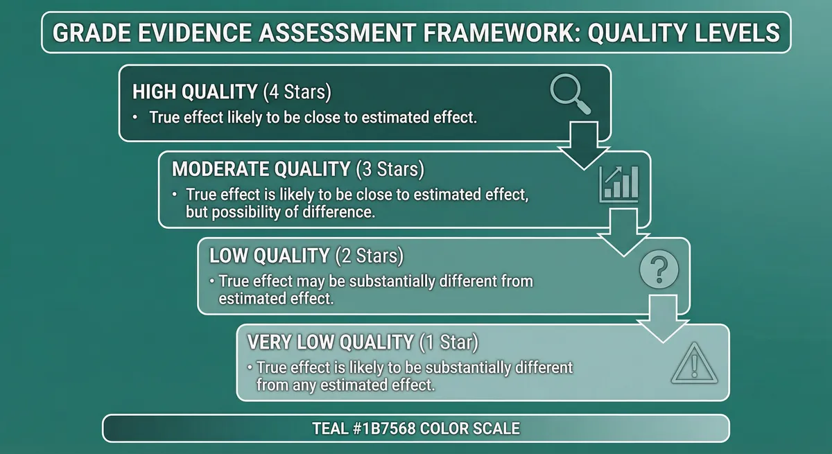
GRADE Evidence Level: Moderate
RCTs with limitations or strong observational studies
Microsurgical flap salvage and tissue replantation represent the most extensively documented clinical application of medicinal leech therapy and the basis for the organism's 2004 FDA clearance. This page presents the complete outcome data for this indication: 4 systematic reviews encompassing over 400 cases, body-region-specific evidence for digit, ear, and breast replantation, institutional protocols from leading U.S. academic medical centers, cost-effectiveness analysis, and thorough Aeromonas infection management data. The evidence consistently demonstrates a 78% overall salvage rate across multiple independent reviews, with outcomes critically dependent on timing of intervention, prophylactic antibiotic adherence, and institutional protocol standardization.
FDA 510(k) Clearance Status
510(k) Clearances for Medicinal Leeches
| 510(k) Number | Year | Applicant | Annual U.S. Supply (est.) |
|---|---|---|---|
| K040187 | 2004 | Ricarimpex SAS (France; 150+ years breeding history) | see FDA AccessData |
| K132958 | 2014 | Biopharm (UK) Ltd. (via Carolina Biological Supply Co.) | see FDA AccessData |
2024 Regulatory Transfer
Outcome Metrics at a Glance
78%
Overall Salvage Rate
Confirmed by two independent systematic reviews (Whitaker 2012; Kuhn 2019)
92%
Prophylactic Salvage
vs 67% reactive (p<0.05) in digit replantation
14.4%
Infection Rate
<5% with prophylaxis; 0% in Nguyen et al. 2012
49.75%
Transfusion Rate
Serial hematocrit monitoring required every 4-8 hours
Systematic Reviews: Converging Evidence
The evidence for leech-mediated flap salvage derives predominantly from systematic reviews of case series — a design limitation imposed by the clinical impossibility of randomizing patients with failing flaps to leech therapy versus no intervention. Withholding an FDA-cleared intervention from a patient with an acutely failing flap would be ethically untenable. Despite this constraint, the convergence of four independent systematic reviews on a salvage rate of 65-83% provides solid evidence for clinical practice.
| Study | Design | Population (n=) | Intervention | Key Outcome | Result |
|---|---|---|---|---|---|
| Whitaker et al. 2012 | Systematic review | Plastic and reconstructive surgery patients with venous congestion across 67 publications (1966-2009) (n=277) | Medicinal leech therapy for flap salvage and tissue replantation | Overall tissue salvage rate, complication profile | 78% salvage (216/277); 88.3% without infection vs 37.4% with infection Landmark review. 49.75% transfusion rate; 14.4% infection rate; 21.8% overall complication rate. Male:female ratio ~2:1; age range 2-81 years |
| Herlin et al. 2017 | Systematic review + retrospective cohort | Free flap patients with venous congestion (n=4) | Medicinal leeches with ciprofloxacin + TMP-SMX prophylaxis; 41 studies reviewed + own 43-patient series | Flap salvage rate by timing of intervention | 83.7% salvage within 24 hours of congestion onset; 38.6% when delayed beyond 24 hours 45-percentage-point decline with delayed therapy. Recommended cipro + TMP-SMX as first-line prophylaxis. Optimal frequency: every 2-8 hours; duration: 4-10 days |
| Kuhn et al. 2019 | Comprehensive review | All plastic and reconstructive surgery flap patients (n=NR) | Medicinal leech therapy as standard of care for venous decompression | Confirmation of salvage rate and standard-of-care positioning | 78% salvage confirmed; positioned leech therapy as standard of care when surgical revision fails Published in Plastic and Reconstructive Surgery — Global Open. Confirmed FDA 510(k) clearance since 2004 |
| Smolle et al. 2024 | Systematic review | Breast reconstruction patients (75% reconstructive, 25% other breast surgery); 18 studies (4 case series, 14 case reports) (n=4) | Medicinal leech therapy for flap congestion; median 2 leeches/session, 3 sessions/day, 3 days duration | Tissue salvage rate and complication burden | 75% salvage; 81% complication rate (infection and anemia) First systematic review dedicated to breast surgery. Concluded leech therapy must be judiciously used given high complication burden |
Key Convergence Points
Three principal systematic reviews converge on a salvage rate of approximately 78%. Whitaker et al. (2012) established this benchmark across 277 cases and 67 publications spanning four decades. Kuhn et al. (2019) independently confirmed the 78% rate and positioned leech therapy as the standard of care when surgical revision fails or is not technically feasible. Herlin et al. (2017) demonstrated that timing is the dominant prognostic variable: 83.7% salvage within 24 hours of congestion onset versus only 38.6% beyond 24 hours — a 45-percentage-point decline that highlights the urgency of early intervention.
Smolle et al. (2024) contributed the first body-region-specific systematic review for breast surgery, documenting a 75% salvage rate with an 81% complication rate — reflecting the greater complexity of large-volume flap management compared to digits and ears.
Herlin et al. drew an important mechanistic distinction: leech therapy is effective for intrinsic venous insufficiency within the flap (microthrombi, inadequate venous outflow) but is unlikely to salvage a flap with proximal venous anastomosis or pedicle obstruction , which requires surgical revision.
Evidence Level Classification
Timing and Application Strategy
Time Is Tissue
Pathophysiology of Venous Congestion
- Venous obstruction or insufficiency from thrombosis, kinking, vasospasm, or inadequate anastomosis
- Blood accumulates in tissue; interstitial pressure rises
- Capillary perfusion ceases as arterial inflow is impeded by elevated venous back-pressure
- Tissue ischemia and edema develop; flap becomes indurated and cyanotic
- Microthrombi cascade in small vessels
- Irreversible necrosis occurs within 6-8 hours without intervention
1. Active Blood Extraction
Each leech ingests 5-15 mL during a 30-90 minute feeding, creating immediate mechanical decompression. The negative pressure from peripharyngeal muscle contraction actively draws venous blood from the congested tissue bed.
| Study | Design | Population (n=) | Intervention | Key Outcome | Result |
|---|---|---|---|---|---|
| Soucacos et al. 1994 | Case series | Replanted fingers/hands (29 patients) and free tissue transfers (18 patients) (n=29) | Medicinal leech therapy for venous insufficiency in replantation and tissue transfer | Resolution of venous insufficiency | 24/29 replantation cases and 18/18 tissue transfer cases salvaged (83% replantation; 100% free tissue transfer) One of the largest single-center series for digit/hand replantation |
| Hayden et al. 1988 | Animal experimental (porcine model) | Sacral skin flaps with induced venous occlusion in pigs (n=NR) | Medicinal leech application with laser Doppler blood flow monitoring | Blood flow restoration in congested flaps | Substantial blood flow increase within 1 hour in all 9 congested cases Preclinical confirmation of the microcirculation improvement mechanism. Established the experimental foundation for clinical use |
| Uncontrolled report (unverifiable) 1998 | Controlled study | Pharynx/esophagus defects (98 patients) and auricular injuries (12 patients) (n=98) | Pre- and postoperative hirudotherapy with interstitial polarography monitoring | Tissue oxygen restoration and transplant viability | Oxygen restored in 69.9%; relative normal in 30.2%. Functional outcomes improved 26.2-48.4% vs controls Among the largest published series. Marginal flap necrosis in only 12-30%. Transplant viability preserved in all cases |
| Nguyen et al. 2012 | Case series | Native skin (5), local flaps (6), regional flaps (14), free flaps (14) (n=NR) | Medicinal leech therapy with prophylactic antibiotics for venous congestion | Salvage rate by flap type; Aeromonas infection rate with prophylaxis | 100% salvage (native/local); 33% salvaged, 33% partial, 33% lost (regional/free). 0% Aeromonas infections Salvaged group required significantly fewer leeches (38 vs 158). Prophylactic antibiotics eliminated Aeromonas infections entirely |
Impact of Infection on Salvage
Whitaker et al. (2012) demonstrated that Aeromonas{" "} infection catastrophically reduces salvage outcomes:{" "} 88.3% salvage without infection vs 37.4% with infection {" "} — a 51-percentage-point decline. This single finding justifies the mandatory antibiotic prophylaxis protocols now universally recommended. Infection prevention is the most impactful modifiable factor in treatment outcomes.
Leech Consumption as Prognostic Indicator
Nguyen et al. (2012) found that the salvaged group required significantly fewer leeches (38 +/- 34 mean) than the lost group (158 +/- 224 mean), suggesting that{" "} early favorable response is a prognostic indicator. High leech consumption without clinical improvement within 48 hours should prompt reassessment of the diagnosis, including evaluation for proximal venous obstruction requiring surgical revision.
Response Criteria
Digit Replantation Evidence
FDA-Cleared Indication
Digital replantation with venous congestion falls within the FDA 510(k)-cleared indication for venous decompression. Prophylactic leeching achieves 92% digit survival versus 67% with reactive application (p<0.05).
Distal fingertip amputation at or distal to the distal interphalangeal joint presents a unique microsurgical problem. At this level, veins are typically too small (0.3-0.5 mm) for reliable anastomosis. Even when venous repair is attempted, thrombosis rates are high. The result is a replanted digit with arterial inflow but inadequate venous outflow — the precise indication for which the FDA cleared medicinal leeches in 2004. Before the introduction of leech therapy, the salvage rate for distal fingertip amputations was dismal. Leeches transformed this indication from a near-certainty of failure to a procedure with realistic salvage expectations.
| Study | Design | Population (n=) | Intervention | Key Outcome | Result |
|---|---|---|---|---|---|
| Prophylactic vs. reactive study 2020 | Retrospective cohort | Distal digit replantation patients (n=NR) | Prophylactic leeching (before congestion develops) vs reactive leeching (after congestion is clinically apparent) | Complete digit survival and early complication rate | 92% survival (prophylactic) vs 67% survival (reactive); p<0.05 Early complications 8% (prophylactic) vs 50% (reactive). 25-percentage-point improvement represents a paradigm shift toward prophylactic protocol |
| Soucacos et al. 1994 | Case series | Replanted fingers/hands (29 patients) with venous insufficiency (n=29) | Medicinal leech therapy for venous congestion in digit/hand replantation | Digit/hand salvage rate | 24/29 cases salvaged (83%) One of the largest single-center digit replantation series with leech therapy |
| De Sena et al. 2019 | Retrospective cohort | Digit revascularization and replantation patients (n=NR) | Medicinal leech therapy with variable duration; identified 4.5-day threshold | Digit survival by leeching duration | Digits leeched >4.5 days had significantly higher survival rates Longer leeching associated with higher transfusion incidence and longer hospital stay. Important for cost-efficiency calculations |
| Brody, Maloney & Hentz 1989 | Case series | Digit replantation patients with relative contraindications for procedure (n=NR) | Medicinal leech therapy when venous repair was technically impossible | Digit salvage; infection and transfusion rates | All digits salvaged; no infections; no transfusions required Unusually favorable complication profile. Demonstrated feasibility in high-risk patients with relative contraindications |
| Barnett 1998 | Case series | Complete fingertip amputations distal to the DIP joint (n=NR) | Artery-only replantation with medicinal leech therapy for venous decompression | Fingertip salvage in amputations where venous repair was impossible | Successful replantation in majority of cases; leech therapy sustained venous drainage until neovascularization Demonstrated viability of artery-only replantation approach for distal amputations |
Prophylactic vs Reactive Leeching
A retrospective study of 25 digits (12 reactive, 13 prophylactic) demonstrated a{" "} 25-percentage-point improvement in survival {" "} (67% to 92%) and dramatic reduction in early complications (50% to 8%) with prophylactic leeching. The implication: in distal digit replantation where venous anastomosis is absent or unreliable, prophylactic leech application (2-3 times daily from the early postoperative period) should be initiated as part of the standard postoperative protocol rather than waiting for congestion to develop.
| Parameter | Frequency | Action Threshold |
|---|---|---|
| Hemoglobin/Hematocrit | Every 4 hours during active leeching | Transfuse pRBC if Hgb <7 g/dL (<8 g/dL with cardiac comorbidity) |
| Platelet count | Every 8 hours | Platelet transfusion if <50,000/µL with active bleeding |
| Coagulation panel (PT/INR, aPTT) | Daily | Correct coagulopathy if bleeding is excessive |
| Type and crossmatch | On admission | Maintain 2 units pRBC available at all times |
| Fluid balance | Continuous | Aggressive IV hydration to compensate for blood loss |
Ear Replantation Evidence
FDA-Cleared Indication
Ear replantation with venous congestion falls within the FDA 510(k)-cleared indication. Artery-only replantation with leech-assisted venous decompression achieves approximately 80% salvage — a procedure previously considered unfeasible without microvascular venous anastomosis.
Total ear amputation is a devastating injury. The auricle's complex three-dimensional architecture — thin cartilaginous framework covered by delicate skin — makes reconstruction with prosthetics or autologous tissue grafts aesthetically inferior to replantation. Yet microsurgical ear replantation remains among the most technically demanding procedures in plastic surgery: vessels are small (0.3-0.7 mm), vessel identification is difficult, and the distinction between arteries and veins can be impossible in the traumatized field. Over 84 ear replantations have been described in the published literature since Pennington's first successful microsurgical ear replantation in Sydney in 1980.
The accumulated evidence supports a significant finding: artery-only replantation with medicinal leech therapy for venous decompression is a viable clinical approach when venous repair is technically impossible. Surgeons should not abandon ear replantation attempts solely because venous outflow cannot be established microsurgically.
| Study | Design | Population (n=) | Intervention | Key Outcome | Result |
|---|---|---|---|---|---|
| Cho (aggregate analysis) 2016 | Literature analysis | Total ear replantation cases from published literature (n=NR) | Microsurgical replantation with leech-assisted venous decompression | Overall survival; salvage rate after venous congestion | ~68% overall survival; 80% salvage rate with leeches after venous congestion; 57% required external decompression Significant finding: artery-only replantation with leech decompression demonstrated as viable clinical approach |
| Concannon & Puckett 1999 | Case report | Partial ear amputation (n=NR) | Microsurgical partial ear replantation with medicinal leech therapy for venous decompression | Ear salvage | Successful replantation with leech-assisted venous drainage Published in Annals of Plastic Surgery |
| Cho & Bhangoo 1998 | Case report | Pediatric patient (child) with complete ear amputation (n=NR) | Microsurgical ear replantation without venous repair; leeches for venous decompression | Ear salvage without venous anastomosis | Successful replantation. Third reported case of ear replantation in a child without venous repair Published in British Journal of Plastic Surgery. Demonstrated pediatric applicability |
| Kind et al. 2001 | Case report | Complete ear amputation (n=NR) | Complete ear replantation without venous anastomosis; leech therapy for venous decompression | Ear salvage with artery-only technique | Successful complete ear replantation relying entirely on leech therapy for venous drainage Published in Microsurgery. Reinforced viability of artery-only approach |
| Mutimer, Banis & Upton 1987 | Case series | Total ear amputation patients; vessel identification difficult in traumatized field (n=NR) | Ear reattachment with leech therapy when heparin and plasminogen activators failed | Ear salvage and microcirculation restoration | Successful salvage in cases where pharmacologic approaches (heparin, plasminogen activators) failed Landmark pediatric case of 5-year-old boy demonstrated leech efficacy in children |
Clinical Algorithm for Traumatic Ear Amputation
- Attempt microsurgical replantation — arterial and, if possible, venous anastomosis
- If venous anastomosis fails or is impossible — proceed with arterial-only replantation
- Initiate leech therapy immediately in the early postoperative period for venous decompression
- Continue leech therapy for 2-10 days until neovascularization establishes collateral venous drainage
- Monitor hematologic parameters closely; transfuse as indicated
Expected salvage rate with this approach: approximately 80%.
Systems-Level Prevention
Decision Algorithm for Breast Reconstruction Flap Congestion
- Identify congestion: Dark color, turgor, brisk dark capillary refill, tissue oximetry <30%
- Immediate surgical exploration: Rule out pedicle kinking, compression, or thrombosis
- If surgically correctable: Revise anastomosis or augment venous outflow with supplementary vein
- If not surgically correctable: Initiate leech therapy
- Monitor response at 4-6 hours: If no improvement, re-explore surgically
- Continue if responding: Up to 5-7 days with close hematologic monitoring
- Accept partial salvage: In large-volume flaps, partial necrosis with preservation of sufficient tissue volume may still achieve acceptable reconstruction
Institutional Protocols: U.S. Academic Medical Centers
The implementation of leech therapy in U.S. hospitals has matured from ad hoc bedside application to standardized, protocol-driven care. Three institutional models merit specific discussion as frameworks for implementation.
University of Iowa — Head and Neck Protocol
- Leeches applied every 2 hours for acutely congested free flaps
- Serial hematocrits obtained every 8 hours throughout treatment
- Concomitant systemic anticoagulation per microsurgical flap protocol
- Dedicated nursing assessment of flap perfusion between leech applications
- Immediate surgical re-exploration if perfusion does not improve within 4-6 hours
| Assessment | Frequency | Action Thresholds |
|---|---|---|
| Flap color, turgor, capillary refill | Every 1-2 hours | Dark purple/turgid: apply leeches; pale/cool: evaluate arterial inflow |
| Tissue oximetry (if available) | Continuous | SpO&sub2; <30%: congestion; <20%: impending failure |
| Hemoglobin/hematocrit | Every 4-8 hours | Transfuse if Hgb <7 g/dL (or <8 g/dL in cardiac comorbidity) |
| Temperature at bite site | With each assessment | Rising temperature may indicate infection |
| Wound culture | If signs of infection | Start empiric treatment; adjust based on culture sensitivities |
Cost-Effectiveness Analysis
Published cost analyses provide a framework for evidence-based resource allocation in leech therapy. Individual leeches cost approximately $10-15 each from FDA-cleared suppliers. A typical course uses 30-160+ leeches depending on tissue type and duration, translating to a direct leech cost of $300-$2,400 per patient — a fraction of the total hospitalization cost ($12,622-$123,563).
| Study | Design | Population (n=) | Intervention | Key Outcome | Result |
|---|---|---|---|---|---|
| Cost-efficiency study 2022 | Cost-effectiveness analysis | Revascularized and replanted digits with venous congestion (n=NR) | Medicinal leech therapy; incremental cost per additional day: $2,951 | Cost-efficiency thresholds using $100,000/QALY benchmark | Cost-efficient through 3 days (single digit), 5 days (thumb), 7 days (multiple digits) Hospitalization cost range: $12,622-$123,563. Published in Plastic and Reconstructive Surgery — Global Open |
| De Sena et al. 2019 | Retrospective cohort with cost analysis | Digit revascularization and replantation patients (n=NR) | Leech therapy duration analysis with associated resource use | Survival rate by therapy duration; transfusion and hospitalization costs | Success rate: 69% (1-3 days), 42% (4-7 days), 8% (>7 days) Longer leeching increases survival probability but with diminishing returns and escalating costs |
| Nguyen et al. 2012 | Case series with resource analysis | 39 patients with venous congestion across flap types (n=39) | Leech therapy with quantification of leech consumption by outcome group | Leech use as prognostic indicator | Salvaged group: 38.3 leeches (mean); lost group: 157.9 leeches (mean). 57.9% transfusion rate Lower leech consumption correlates with favorable prognosis, suggesting early response predicts outcome |
Cost-Efficiency Thresholds
Using the standard $100,000/QALY benchmark at{" "} $2,951 per additional day of leech therapy:
| Indication | Cost-Efficient Through |
|---|---|
| Single digit | 3 days |
| Thumb | 5 days |
| Multiple digits | 7 days |
Clinical Interpretation
Aeromonas Infection Management
The Primary Complication: <em>Aeromonas hydrophila</em>
| Prophylaxis Status | Infection Rate | Source |
|---|---|---|
| No prophylaxis | 7-20% | Aggregate literature |
| With appropriate prophylaxis | <5% | Aggregate literature |
| With prophylaxis (Nguyen et al.) | 0% (0/39) | Nguyen et al. 2012 |
| Without optimized prophylaxis (Palm et al.) | Infections occurred in this group only | Palm et al. 2022 |
| Study | Design | Population (n=) | Intervention | Key Outcome | Result |
|---|---|---|---|---|---|
| Lineaweaver et al. 1992 | Multicenter case series | Replantation and flap surgery patients with Aeromonas infection (n=NR) | Documentation of A. hydrophila infections following leech therapy | Infection characteristics, timing, and severity | Onset 24 hours to >10 days post-application; severity from minor wound complications to tissue loss and sepsis Seminal study that established the mandate for routine antibiotic prophylaxis during leech therapy |
| Palm et al. 2022 | Quality improvement study | Patients receiving leech therapy at a tertiary academic medical center (n=NR) | Standardized EMR order panel linking leech orders to automatic antibiotic prophylaxis | Infection rate with vs without optimized prophylaxis protocol | Infections occurred exclusively in subset without optimized prophylaxis; zero infections with EMR-linked protocol Systems-level intervention. Panel includes leech order + antibiotics + labs + nursing protocol + pharmacy notification |
| Nguyen et al. 2012 | Case series | 39 patients receiving leech therapy with prophylactic antibiotics (n=39) | Prophylactic antibiotic coverage throughout leech therapy course | <em>Aeromonas hydrophila</em> infection rate | 0% Aeromonas infections (0/39) Demonstrates that appropriate prophylaxis can eliminate clinical Aeromonas infections entirely |
| Giltner et al. 2013 | Case report | Patient with cellulitis despite ciprofloxacin prophylaxis (n=NR) | Ciprofloxacin monoprophylaxis during leech therapy | Breakthrough Aeromonas infection | Ciprofloxacin-resistant A. hydrophila cellulitis developed despite prophylaxis Published in Journal of Clinical Microbiology. Early warning of emerging ciprofloxacin resistance |
| Wilmer et al. 2014 | Case report | Digit replantation patients with resistant Aeromonas infection (n=NR) | Leech therapy for digit replantation with ciprofloxacin prophylaxis | Ciprofloxacin-resistant infection in digit replantation | 2 cases of ciprofloxacin-resistant A. hydrophila infection complicating digit replantation Published in Journal of Hand Surgery. Reinforced need for dual-agent prophylaxis |
Antibiotic Prophylaxis Protocol
Recommended Dual-Agent Prophylaxis
| Agent | Dose | Notes |
|---|---|---|
| Ciprofloxacin (first-line) | 500 mg PO BID | Historically the standard; rising resistance noted |
| Trimethoprim-sulfamethoxazole (TMP-SMX) | 160/800 mg PO BID | Combination agent; addresses resistance gap |
| Alternative: ceftriaxone | 1 g IV daily | Third-generation cephalosporin; used at some centers |
The Ciprofloxacin Resistance Crisis
Proactive Batch Surveillance Protocol
An innovative approach to resistance management developed following a multidrug-resistant case (IDCases 2020):
- On delivery: Sacrifice one leech per batch of 50 for culture and sensitivity testing
- Every 30 days: Repeat culture of one leech from each stored batch
- Prophylaxis: Directed toward the susceptibility profile of the batch in use
- Quarantine: Batches harboring multidrug-resistant isolates are quarantined and not used for patient care
Result: No further leech-associated infections after protocol implementation.
Immunocompromised Patients
Comparison with Alternative Venous Decompression Methods
Leeches are not the only option for venous decompression, though they remain the standard of care. A 2011 systematic review of chemical and mechanical alternatives concluded that no single alternative has demonstrated superiority over medicinal leeches in controlled comparisons.
| Method | Advantages | Limitations |
|---|---|---|
| Medicinal leeches (standard of care) | Combined mechanical + pharmacologic effect; 78% salvage; FDA 510(k)-cleared | Infection risk (Aeromonas); transfusion requirements; patient aversion |
| Heparin pledgets / chemical leech | Available; easy application; local action | Less effective for free flaps; no pharmacologic synergy |
| Subcutaneous heparin + scarification | No infection risk | Requires repeated scarification; less blood removal |
| Negative pressure wound therapy (NPWT) | No infection risk; continuous drainage | No anticoagulant effect; limited data |
| Hyperbaric oxygen therapy | May reduce ischemia-reperfusion injury | Expensive; logistically difficult; adjunctive only |
Evidence Summary and GRADE Assessment
| Indication | Best Available Evidence | Level | FDA Status |
|---|---|---|---|
| Venous congestion in flaps/replants | Systematic reviews (Whitaker 2012, Herlin 2017, Kuhn 2019) | IV (SR) | FDA 510(k)-cleared |
| Digit replantation | Case series, retrospective studies | IV | FDA 510(k)-cleared |
| Ear replantation | Case reports/series (>84 cases cumulative) | IV | FDA 510(k)-cleared |
| Breast reconstruction flap salvage | Systematic review (Smolle 2024) | IV (SR) | FDA 510(k)-cleared |
| Head and neck free flap reconstruction | Uncontrolled Russian-language report, not independently verifiable | III | FDA 510(k)-cleared |
Why No Randomized Controlled Trials Exist
Evidence Gaps and Research Priorities
Multicenter Prospective Registries
Standardized data collection across microsurgical centers with defined endpoints (salvage rate, time to intervention, hematologic parameters, infection rate, resistance profiles) would strengthen the evidence base beyond retrospective case series.
Related Research
Leech Therapy For The Treatment Of Venous Congestion In Flaps, Digital Re-Plants And Revascularizations - A Two-Year Review From A Regional Centre
Two-year regional review of leech therapy in 18 patients (20 tissues): 65% successful tissue salvage with 100% success in pedicled flaps, 67% in digital revascularizations, 25% in replants and 0% in free flaps.
Butt AM et al. · Journal of Ayub Medical College, Abbottabad
The use of medicinal leeches, Hirudo medicinalis, to restore venous circulation in trauma and reconstructive microsurgery
In 24 of 29 patients, venous insufficiency after replantation of digits/hands or microsurgical free tissue transfer was successfully treated with medicinal leeches, demonstrating leech therapy as a valuable alternative to surgical revision of venous outflow.
Soucacos PN, Beris AE, Malizos KN et al. · International angiology
Endodontic microsurgery virtual reality simulation and digital workflow process in a teaching environment
Computer simulations are stimulating increased attention in dentistry.
Carpegna G et al. · European journal of dental education : official journal of the Association for Dental Education in Europe
Near-Infrared Spectroscopy for Continuous Noninvasive Monitoring of Free Flap in Head and Neck Reconstruction: Systematic Review of the Literature and Personal Experience
Buried free flaps represent a reconstructive challenge concerning monitoring of vitality, which is fundamental for an early detection of flap failure and prompt surgical salvage.
Festa BM et al. · Surgical innovation
Leech Therapy Following Digital Replantation and Revascularization
Venous congestion after digital replantation or revascularization threatens digit survival in the immediate postoperative period. External bloodletting, including leech therapy, provides a central role in salvage of the congested finger.
Pickrell B et al. · The Journal of hand surgery
A global view of digital replantation and revascularization
Global review of digital replantation/revascularization survival rates across regions. Discusses multi-team protocols, flow-through and pedicled flaps for multi-digit and total-hand replantations.
Tang JB et al. · Clinics in plastic surgery